LAB 9 :THE SYSTEMIC CIRCULATORY SYSTEM

Objectives:
1)Understand the general function of the arterial, venous, and lymphatic systems.
2)Understand the function of portal systems.
3)For each animal, learn the major blood vessels that are discussed in this lab manual.
4)For each animal, learn the pathway of blood flow through the body.
THE CIRCULATORY SYSTEM
In this lab we will be looking at the circulatory system in the pleuroperitoneal cavity of the dogfish and mud puppy and in the abdominal cavity of the mammal. This system basically consists of the systemic arterial branches of the dorsal aorta, the renal (kidney) and hepatic (liver) portal systems and the systemic venous system leading from the body to the heart. In addition, we will look at the lymphatic system.
The portal systems transport venous blood from one organ to a different part of the same organ or a different organ.
Useful terms: celiac (celiac) = bellygastero = stomach phrenic = diaphragm
lieno = spleenhepatic = liverrenal = kidney
Water and ion exchange into tissues at the arterial end of capillary beds is greater than the exchange into the capillaries at the venous end. As a result, there is a net loss from the blood. The lymphatic system evolved to get excess water back into the circulatory system. Nodes and various organs and glands associated with this system tend to be involved in immune responses.
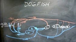
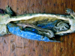
DOGFISH CIRCULATORY SYSTEM
Arterial System
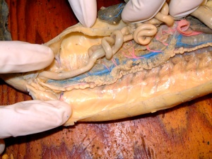
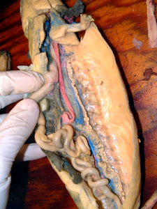
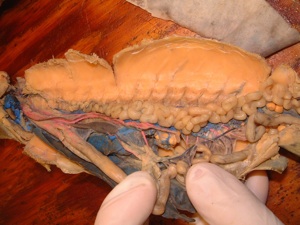
The arterial system in the dogfish is injected in red. Last week you looked at the efferent arteries leading from the gills and the paired dorsal aorta and internal carotids leading to the head. The coronary arteries leave the second and third gill loops, then join, to take blood to the heart. The efferent arteries join to form the dorsal aorta, which enters the pleuroperitoneal cavity. We will look at the somatic branches of the dorsal aorta first. Find the first paired branch, the subclavians, which arise just anterior to the fourth efferent artery and serve the pectoral fins. The dorsal aorta gives rise to many intersegmental arteries, which carry blood to the segmented muscles and form renal arteries to the kidneys. Posteriorly, the dorsal aorta gives rise to the paired iliac arteries leading to the pelvic fins as the femoral arteries. The dorsal aorta continues in the tail as the caudal artery.
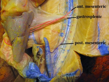
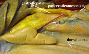
The visceral branches of the dorsal aorta may be paired or unpaired. There are four major branches off of the dorsal aorta. The first is the celiac artery, which departs above the liver to run in the mesentery and go to several organs. The branches indicate the organs they serve. The first branch is genital (ovarian or testicular), then the gastrohepatic and pancreaticomesenteric, the duodenal and anterior intestinal arteries. The next artery leaving the dorsal aorta is near the spleen. The anterior mesenteric artery goes to the intestine and its valves. Just posterior to it is the gastrosplenic artery to the spleen, stomach and dorsal lobe of the pancreas. A little ways posterior to this branch is the posterior mesenteric artery leading to the rectal gland.
Venous Systems
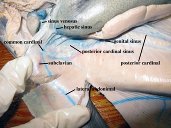
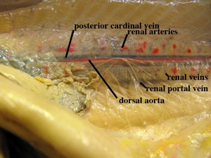
The renal portal system takes the blood from the caudal vein in the tail to the kidneys via the two renal portal veins. The blood enters the kidneys on afferent renal veins, which connect, via capillaries to efferent renal veins, which drain the kidneys into the posterior cardinal veins and then to the posterior cardinal sinuses.
The hepatic portal system takes the blood from the abdominal viscera to the liver. In the dogfish, this system is injected yellow. The liver can metabolize, detoxify and store substances in the blood. Branches from the organs (gastric, lienomesenteric, pancreaticomesenteric veins) come together to form the main hepatic portal vein, which enters the liver and subdivides. From the liver, blood is drained via the hepatic veins of the systemic venous system to the sinus venosus.
Systemic veins usually run close to the arteries and share the same name. These are injected blue or not injected at all. Sometimes larger pools of venous blood are found in sinuses. We have already examined the renal portal system. The posterior cardinal veins drain into the posterior cardinal sinuses, which also connect with the genital sinuses. The posterior cardinal sinuses drain into the common cardinal veins and then the sinus venosus of the heart. Posteriorly the femoral, iliac and cloacal veins enter the lateral abdominal veins located in the lateral muscles of the body wall. Intersegmental veins join in, as do the subclavian veins just before they all enter the common cardinal veins and sinus venosus. The head is drained by sinuses, which enter via the anterior cardinal sinuses. The anterior cardinal sinuses are deep in the head behind the eye and will not be observed here. The small jugular veins also enter the common cardinal sinuses, which then lead to the sinus venosus and ventricle of the heart. The jugular veins are deep in the lower jaw, so they will not be observed.
Lymphatic System
In the dogfish, a number of sinusoids drain extra circulatory fluids into the veins, and appear to function like the lymph system of other vertebrates. A large spleen is also present.
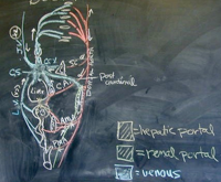
NECTURUS CIRCULATORY SYSTEM
Arterial System
Just after the junction of the two radices (aortic roots) of the dorsal aorta, the two subclavian arteries branch off to serve the arms. Cutaneous (lateral) arteries branch off, penetrate the muscles, and run just under the skin along the horizontal septum, where they serve the skin and muscles. Note the pulmonary artery runs the length of each lung after leaving the junction of the second and third efferent arteries. The dorsal aorta gives off intersegmental arteries. Some of these supply the kidneys (renal arteries), gonads (genital arteries), and the hind limbs (iliac and femoral arteries). The dorsal aorta continues to the tail as the caudal artery. The ventral arteries coming off the dorsal aorta are the single gastric artery taking blood to the stomach, which can be found just posterior to the subclavians. Just posterior to the gastric artery is the single, celiacomesenteric artery with branches to the organs (splenic, hepatic, pancreaticoduodenal, mesenteric). Posteriorly, a number of mesenteric arteries go to the large and small intestines. Note that this system shows little change from that of a dogfish, although the celiac and mesenteric arteries are joined.
Venous System
The renal portal system is more primitive than that of the dogfish in that the embryonic connection of the posterior cardinal veins to the sinus venosus is not lost anterior to the kidney. The blood from the tail and hind limbs is taken to the kidneys via the afferent renal portal veins, which run on the lateral (outside) edge of the kidneys. After passing through the kidney, blood is collected in the efferent renal veins into the posterior vena cava and taken to the sunus venosus of the heart.
The hepatic portal system is injected yellow in the mud puppy. Venous blood enters the liver from branches that drain the tail and hind limbs (abdominal vein), intestine (mesenteric vein), gastrosplenic vein and pancreaticoduodenal vein. Look in the mesentery below the intestine to see the but dark or yellow injected mesenteric vein. The major change that has taken place is that now some of the blood from the hind limbs, cloaca, bladder and body wall enters the hepatic portal system via the ventral abdominal vein. This vein is homologous to the lateral abdominal veins of the shark, which now forms a single vein and lies mid-ventrally stuck to the inside of the body wall and is usually dark brown with blood.
The lungs have pulmonary veins along their length, which unite anteriorly to form a single vessel entering the left atrium of the heart. A new, single vein, the posterior vena cava, drains the posterior region of the body. This is partly made up from the embryonic paired posterior cardinal veins and the right hepatic vein. It runs up the midline between the kidneys, receiving the renal veins, then the ovarian or testicular veins, hepatic veins at the liver and anterior to the septum splits into two hepatic sinuses, which join the common cardinal veins.
Since the posterior cardinal veins persist in the mud puppy, some blood from the kidneys (subcardinal veins) and the body wall (inter segmental veins) is drained to the common cardinals via these vessels. The common cardinal veins also receive blood from the anterior of the body. The internal and external jugular veins are deep in the head and the lingual vein is deep in the tongue so they will not be dissected. They drain into the subclavians from the arms and they enter the common cardinal veins. The common cardinal veins, after collecting blood from the posterior cardinal veins, lateral veins, cutaneous veins and posterior vena cava, enters the sinus venosus and right atrium of the heart.
Lymphatic System
In amphibians, there is a well-developed lymphatic system in the subcutaneous area to return fluids to the blood. The lymph flows in channels and is moved by pulsating lymph hearts with valves to eliminate backflow. The spleen is a lymphoid mass.
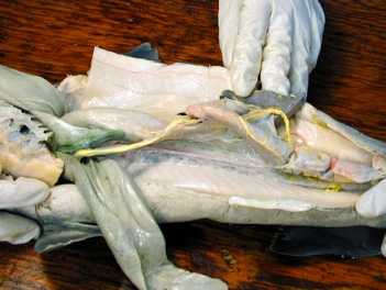
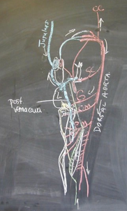
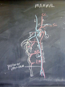
MAMMALIAN CIRCULATORY SYSTEM
Arterial System
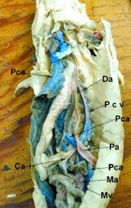
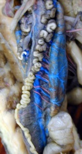
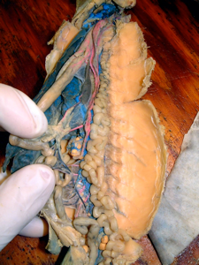
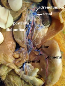
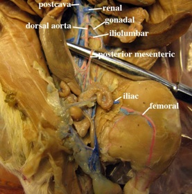
Follow the dorsal aorta into the abdominal cavity. Note the small intercostal arteries and other paired intersegmental arteries, which form the adrenolumbar arteries (to the adrenals), renal arteries (to kidneys), spermatic or ovarian arteries (to gonads). There are many paired, deep, segmental lumbar arteries and one set of paired iliolumbar arteries to the back muscles, as well as iliac and femoral arteries to the limbs. The medial caudal artery down the tail is too deep to be traced. Note in males, the testes have dropped and the gonadal (testicular) arteries (and veins) run parallel to the dorsal aorta to the gonads.
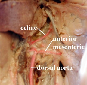
The first unpaired artery, which goes ventrally to the organs, is the celiac artery. It is easier to spot on the right side of the membrane. This artery has many branches; gastric, splenic, hepatic, gastroduodenal are the major ones. The branch to the pancreas joins with the pancreatic branch of the next visceral branch forming a loop. The anterior mesenteric artery lies just posterior to the celiac artery and has branches to the pancreaticoduodenal area (forming the loop), colic and intestinal areas. The posterior mesenteric artery is smaller. It branches to the colon and rectum and then joins with the loop from the anterior mesenteric artery. Note again that this system is similar to that of the dogfish.
Venous Systems
The renal portal system does not exist in mammals.
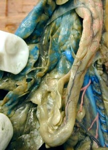
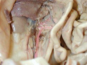
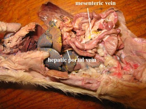
The hepatic portal system is injected (yellow) in your specimen. It drains the viscera from the mesenteric veins, gastrosplenic vein,the stomach, pancreaticoduodenal vein and their branches. The mesenteric vein can sometimes be seen running along the bottom of the dorsal mesentery. They all enter the hepatic portal vein, which leads to the liver along the bile duct. Note that the blood from the hind limbs no longer flows into this system.
Venous blood (injected blue) from the hind limbs flows via the femoral and iliac veins to the single median postcava (posterior vena cava). The caudal vein joins the left common iliac vein. Blood also enters from the iliolumbar and many deep, paired, lumbar veins, the right genital vein (spermatic or ovarian) and the renal veins. The left spermatic or ovarian vein drains via the left renal vein as does the left adrenolumbar. Those on the right side enter separately. Near the diaphragm, the hepatic veins and superior phrenic veins enter the postcava, which then penetrates the diaphragm and enters the right atrium of the heart. In the head region, blood is drained via the external and internal jugular veins. They are in the throat and were observed last week. The subclavian veins from the forelimbs join and together they descend as the innominate (brachiocephalic) veins, joining to form the precava (anterior vena cava). This vessel enters the right atrium of the heart. In rats, the left subclavian and jugular loop under the heart and enter the right atrium via the posterior vena cava.
Lymphatic System
Thin walled, permeable vessels are located throughout the body. Fingerlike capillaries called lacteals project into the villi of the small intestine for fat absorption. These molecules are then transported to the blood. Excess lymphatic fluid is returned to the venous system by a thoracic duct, which opens near the left subclavian vein. Lymph nodes, which filter and destroy viruses and bacteria, are found in the neck, intestinal and groin areas, smaller ones are scattered throughout the system. These nodes also house lymphocytes for immune responses. The thymus gland, on top of the heart, produces lymphocytes. Tonsils, on the palate, act like lymph nodes, eliminating possible foreign invaders. The spleen is engaged in immune response, produces leukocytes, can make erythrocytes and destroy old ones, and is a reservoir for blood platelets.