THE INTEGUMENT AND ITS DERIVATIVES

Objectives:
Describe the components of the integument and their basic functions.
Describe the structures that are produced from keratinization of the epidermis.
Understand the formation and function of glands.
Understand how the dermis contributes to the integument.
Determine the composition of the various types of fish scales.
Understand the composition of teeth and be familiar with different types of tooth disposition, insertion and differentiation
EPIDERMIS AND ITS DERIVATIVES
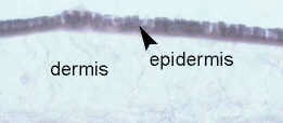
The epidermis, derived from somatic ectoderm, is the exterior-most covering of the chordate body. It provides protection against the invasion of microorganisms, provides flexibility in motion, and seals in moisture. As will be seen, it also gives rise to a variety of differentiated structures such as feathers, hair, horns, claws, nails and glands. Begin by looking at a cross-section of Amphioxus (Branchiostoma) integument. Amphioxus possesses the simplest possible form of epidermis - a single layer of columnar epithelium covered by a thin film of cuticle.
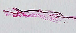
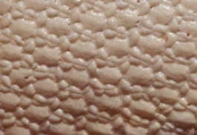
All true vertebrates, however, have developed a multi-layered epithelium. Note the simple, multicellular, epithelium of the lamprey, which has no scales. Fishes and amphibians have a mucus layer for bacterial and mechanical protection and to prevent drying on land. See frog skin.Terrestrial vertebrates have replaced the cuticle with keratin. See the snake skin.
Epidermal Derivatives of the Integument
Keratin Structures
New epidermal cells are formed continuously in the lower layers of the epidermis. In terrestrial vertebrates, new epidermal cells push more superficial ones to the stratum corneum, the outer-most epithelial layer. In the process of self-destruction, these exterior epidermal cells accumulate protein products called keratin. Keratinized or cornified skin serves to prevent water escape and to protect against friction and direct mechanical stimulation (e.g. calluses in humans). The production of all of the following structures involves keratinization:
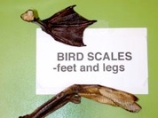
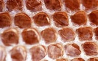
Epidermal Scales: a continuous layer of repetitious thickenings of the stratum corneum; you cannot dissect an individual epidermal scale out of the skin! These scales may be shed entirely (moulting) or in small flakes. Examine preserved specimen of snake skin and dried specimens of bird legs and feet.
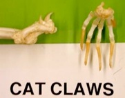
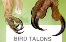
Claws and Talons: curved, laterally compressed keratinized projections from the tips of digits. See dried specimen of cat claws and bird talons.
What are the possible functions of claws and talons?
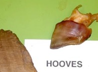
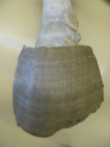
Hooves: enlarged keratinized plates found on the ends of ungulate digits. Examine the hooves of pig and horse.
Nails: keratinized epithelial cells are produced at the nail base and push the existing nail forward. They provide protection from mechanical injury and stabilize skin for better grasping. Found only in primates.
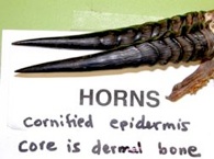
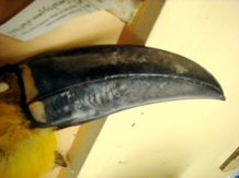
Horns: a tough, cornified layer of the integument covers horns. Their core, however, is bone, which is of dermal origin. Horns are found in bovines (cattle, antelope, sheep, goats, bison, wildebeest). They are retained year-round and grow throughout the animal’s lifetime.
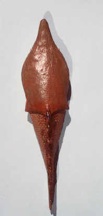
Baleen: found in some whales, baleen is a series of keratinized plates that arise from oral epithelium. These sheets hang from the palate along its length and act as a sieve. See the display.
Of what use would the sieve-like action of these baleen plates be?
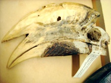
Beaks: epidermal structures, jaws are covered by keratinized sheaths in birds and turtles.
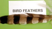
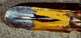
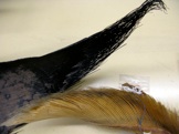
Feathers: are believed to have evolved from reptilian scales. Columns of epidermal cells project into the skin initially to form an invagination called the feather follicle. Later growth results in a projection out of the skin of a keratinized epidermal sheath with an inner feather shaft. These columns then separate and develop into barbs. Feather growth is initiated by dermal papillae, which die in the grown feather to form feather pulp. Examine the dried specimens. Note the quill (calamus), which attaches to the body and extends as a rachis. From the rachis project many veins with barbs and barbules to hold them together.
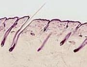
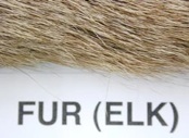
Hair: just as in feathers, there is an initial ingrowth of epidermal cells to form the hair follicle, followed by an outward growth of keratinized cells to form the hair shaft. Dermal papillae cells of the outer edge die and form the core substance of hair follicles. Note the similarities between hair and feathers both in development and in general anatomy. They both possess dermal papillae, shafts, an inner pulp and columns of specialized keratinized cells. Hair is characteristic of mammals.
Which is the living layer of the epidermis in the human scalp?
Which is the non-living layer of the epidermis?
What is the primary function of each epidermal layer?
Glands
Specialized to secrete specific products (oil, sweat, milk, etc.), these structures are derived by an infolding of the epidermis. In many cases they retain a connection to the stratum corneum whereby their secretions can be released at the skin surface.
What do you think is responsible for the slimy feeling of fish skin?
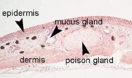
Sketch and label the glands of the frog as seen in the frog integument slide (cross-section).
Sketch and label the human scalp cross-section with oil (sebaceous) and sweat glands.
THE DERMIS AND ITS DERIVATIVES
The dermis is generally much thicker than the epidermis and lies more deeply. It is made of a fibrous mass of connective tissue (collagen) and is of mesodermal origin. It may directly produce dermal (membrane) bone. The dermis is important in defence against injury and in the maintenance of body heat. Deeper regions of the dermis often contain fatty deposits, smooth muscle, blood vessels and nerves. Chromatophore cells are sometimes epidermal, but usually dermal in origin. They secrete melanin, which can be passed to the stratum corneum of skin and to hair shafts to produce colour and block harmful sunlight.
Dermal Bone
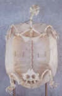
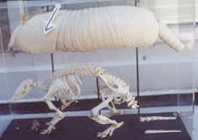
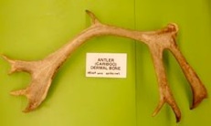
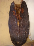
Once present in some extinct fish - Ostracoderms had a complete head shield, while Placoderms had a broken head shield and body armour. Now dermal bone is present in turtle dermal bone, antlers, and in the dermal armour of armadillo. In antlers the velvet is epidermal in origin and shapes and provides blood to the dermal bone. Once grown, the velvet is shed and only the bone remains. Antlers are found in deer, elk, moose and their relatives, often only in males. They are shed annually.
In most modern vertebrates, dermal bone (membrane bone) is formed from embryonic mesenchyme by intramembranous ossification, and contributes to the skull and skeleton, rather than being manifested externally. An exception is teeth, which are partly derived from dermal bone.
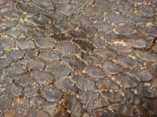
Fish Scales
Fish scales are also called dermal scales since they are derived mainly from the dermis.
-
1)Cosmoid Scales: Found in Placoderms (extinct) as plates, and also typical of the Lobe Finned Fishes or Sarcopterygii, (Choanichthyes). Extinct fish had scales of enamel, cosmine and bone with pulp cavities. Modern ones, like Coelocanth and the lung fish have calcified fibers so this type of scale is almost extinct. No specimens available.
-
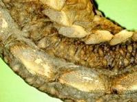
2)Ganoid Scales: See bioplastic mounts, slides, the plates of sturgeon, called scutes, and the scales of the gar pike on display. Made of multi-layered enamel called ganoin over lamellar bone. Primitive (now extinct) species also had a cosmine layer and vascular bone with pulp, but these were lost in modern day examples.
-
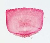
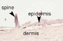
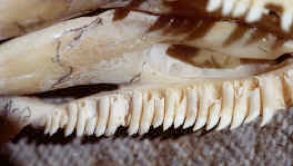
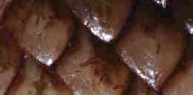
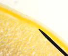
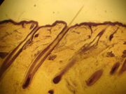
3)Placoid Scales: See bioplastic mounts and dogfish slides. Made of enamel (epidermal) and the dermal derivatives, dentine and bone with a pulp core. They are typical of cartilaginous fishes. Placoid scales are responsible for the rough feeling of dogfish skin.
4) Teleost (bony fish) scales
These are thin scales of dermal bone. They have a thin covering of epidermal tissue over them. It is derived by reduction (loss) of parts of a ganoid scale. There are two types depending on their shape.
4a) Cycloid Scales: See bioplastic mounts and slides. A round ended scale.
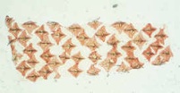
4b) Ctenoid Scales: See bioplastic mounts and slides. A comb shaped end is characteristic of this scale type.
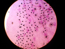
Referring to the bioplastic mount and slides, make a sketch of the placoid, ganoid, cycloid and ctenoid scales. This sketch is for your own reference so do not copy the drawings but draw what you see under the microscope.
TEETH
Teeth are composed of three main parts. Enamel, the hardest substance in the body, covers the tooth surface. It is epidermal in origin. Ganoin is a form of enamel. Dentin is similar to bone in structure but is harder. It is located beneath the enamel and forms the walls of the third component of teeth, the pulp cavity. These are of dermal origin. Cosmine is a form of dentin. Dermal bone called cementum is also present in mammalian teeth.
Teeth are used to catch and hold prey, to crush hard shells and, in some higher vertebrates, to carry out mechanical digestion of food in the mouth.
How does breaking of food into smaller pieces improve its digestion?
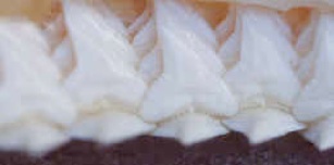
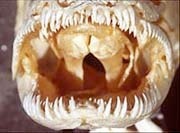
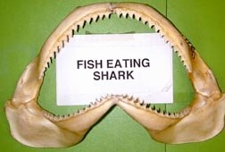
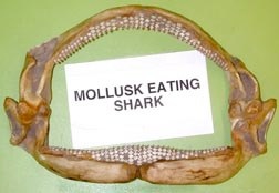
Examine the shark teeth on demonstration. Note that mollusk-eating sharks have blunt teeth, while others have a cutting edge. Despite such variations, the scales formed on dogfish skin and the teeth found in its mouth are both made of enamel, dentin and bone with a pulp core, i.e. shark teeth are modified placoid scales.
Draw both the cross-section and the whole flat-mount of dogfish skin.
Teeth of higher vertebrates are thought to have evolved from bony dermal scales similar to dogfish placoid scales. They have a complex embryonic origin involving both the epidermis and the dermis. Interestingly, their development bears some resemblance to that of hair and feathers. Mesenchyme cells collect in the dermis to form dermal papillae, which are instrumental in the production of dentin and go on to form the pulp of the tooth. Enamel is produced by the epidermis. The tooth in mammals is held in place by cement, which is a non-vascular form of bone.
To compare the various types of dermal scales with teeth, fill in the chart in your lab manual indicating the presence (+) or absence (-) of the indicated substances.
TOOTH CHARACTERIZATION
Tooth Position (where are they found?)
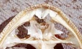
1) Fishes
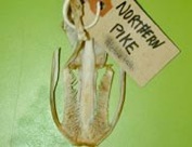
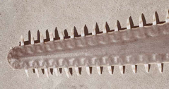
Teeth, or dermal structures, which are tooth-like, may be found wherever ectoderm occurs in the mouth area. For this reason teeth can occur outside the jaws as in the pharyngeal teeth on the bony elements of the branchial bars, or outside the buccal cavity as teeth in the sawfish. See Amia, pike and sawfish.
How does this relate to deuterostome development in vertebrates (recall the development of the mouth and anus)?
2) Amphibians
Teeth occur mainly on the jawbones, with some occurrence on the palate. Examine the skulls of frog (Anurans) and the salamander Necturus (Urodeles) on demonstration.
3) Reptiles
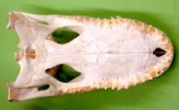
A few reptiles still have teeth on their palate to hold their prey, but most teeth are on the jawbones.
4) Birds
A few birds have teeth on their beaks, but teeth are absent in most birds.
5) Mammals
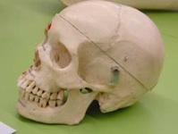
Teeth occur only on the jawbones, i.e. on the dentary, premaxilla and maxilla.
What general trend in tooth location do you observe across the vertebrate phyla?
Tooth Insertion (how they are attached).
There are three methods of tooth insertion. Examine the demonstration material and identify the following methods of insertion:
-
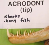
1)Acrodont (acro=end)
The teeth are fused by their bases to the outer surface of the jaw. This condition occurs in most Teleosts (bony fish) and can be also seen in the sharks. The teeth are not firmly rooted and are easily lost and replaced. Teeth that are continually replaced are called polyphyodont (poly=many).
-
2)Pleurodont (pleur=side)
The teeth are fused on one side to the inner surface of the jaw. This occurs in the salamander, Necturus, frogs, and in the lizards. Most birds have no teeth but mergansers, Peking duck and a few others have pleurodont teeth. Pleurodont teeth are usually polyphyodont.
What are reasons for the loss of teeth in most birds?
How do they compensate for this loss?
-
3)Thecodont (theca=cup)
The teeth are placed on the crown of the jaw in a socket. A tooth may have a single root, as in the alligators, or several roots, as in the molars of mammals.
What general trend in tooth insertion do you observe across the vertebrate phyla?
Most thecodont vertebrates replace their teeth only once in their lifetime. Describe some possible reasons for this.
What advantage(s) do acrodonts have over pleurodonts and thecodonts? What disadvantage(s)?
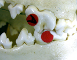
C) Tooth Differentiation (how they differ)
The shape of the teeth in the fishes, amphibia and reptiles is relatively constant in any one group. Functional adaptations occur in size, and in specialization such as poison ducts in the fangs of snakes. Study the demonstration material in the above groups and notice the similarity in shape - called the Isodont or Homodont (iso/homo = equal) condition.
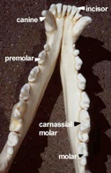
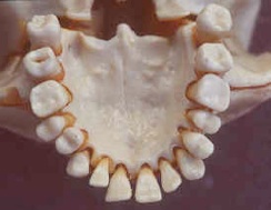
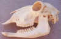
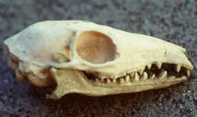
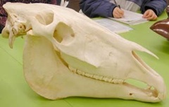
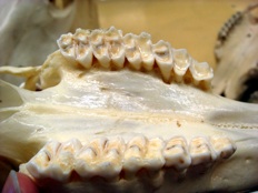
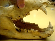
Dentition in mammals is generally Heterodont (hetero=different). The teeth are modified in shape and size to serve specialized functions. Study this modification in the wolf skull. Notice incisors (cutting), canines (piercing), premolars (grinding) and carnassials (shearing), and molars (crushing).
Examine the general demonstration of mammalian teeth. Notice the adaptations are correlated with food habits. Compare the insectivores, herbivores, carnivores and omnivores.
Examine the shrew and mole as examples of insectivores, which eat insects and other small invertebrates. Most of the teeth are generalized and little differentiation has taken place.
Why would insectivores need such long and sharp incisors?
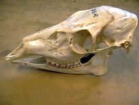
Examine upper and lower jaws of the herbivores, which feed on grasses and other vegetation. Note the differences, a large gap, the diastema (=internal) and often a horny upper pad - check the grazers (horse, deer, ox), and the gnawers (rabbit, beaver).
Why do herbivores have such long jaws formed by the diastema?
Why are the molars and pre-molars so corrugated (alternating ridges and furrows) in herbivores?
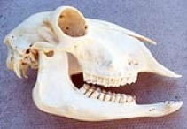
In the carnivores, which feed mainly on herbivores, note the specialized long and sharp canine teeth for biting and piercing, and the last premolar of upper jaw and first molar of lower jaw, which have been modified as carnassials used for cutting. Within the group notice variations of teeth used for cutting, piercing, incising and gripping.
How do you think the carnassials work? (Hint: look at which surfaces touch each other)
Examine the omnivore dentition (pigs, bears, humans) and compare with carnivores, herbivores and insectivores.
Bears are omnivores, eating whatever is available. Note, however, that their overall tooth differentiation resembles that of carnivores. There is a discrepancy here. Why do you think this is so?