Protein Processing II: Endomembrane System -Vesicle Transport and the Golgi
Apparatus
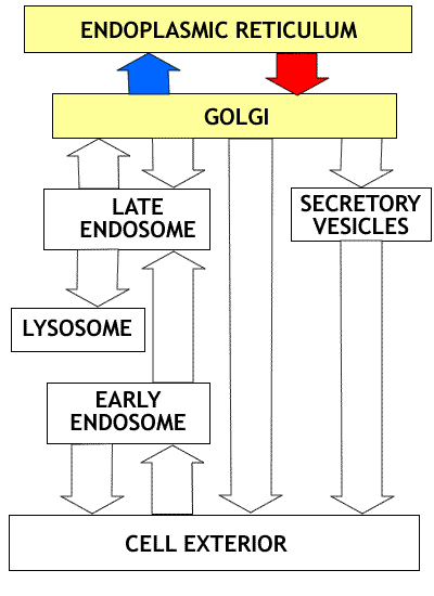 |
In this discussion we will be dealing with
the Endoplasmic Reticulum and the Golgi Apparatus. These compartments of
the endomembrane system are linked by extensive vesicle traffic that moves
proteins in both directions, from ER to Golgi (red arrow), and some proteins
from Golgi to ER (blue arrow). This traffic of proteins, like that occurring
throughout the rest of the endomembrane system occurs by means of vesicle
transport. We start by looking at vesicle transport and then move on to
discussion of the Golgi apparatus. |
Key ideas:
- Vesicles form because of interaction between proteins inserted
into the membrane and special coat forming proteins.
- Formation of the vesicle is an essential for concentration
of cargo proteins.
- Vesicles are targeted by means of protein-protein interaction
(address house number analogy).
- Golgi is the main site of protein processing and sorting
for different destinations.
A. Vesicle Formation and Targeting
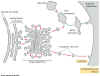
Fig. 14-17 . |
There is a tremendous flux of vesicles within
most cell types. Vesicles form from the
- endoplasmic reticulum, the
- Golgi apparatus and the
- plasma membrane.
They are used to transport membrane and proteins between
many different membranous organelles.
Here we look at how vesicles are formed and how they find
their targets.
|
Vesicle formation and vesicle transport
- Transport between compartments takes place via vesicles.
- Membranes, with both proteins and lipids, and the soluble
proteins contained within the vesicles are transported.
- For example, once the proteins are in the ER, they are transported
by vesicles that bud off of the ER and fuse with the membrane of the
target compartment.
There are two major problems:
- Formation of vesicles and selection of their contents and
- Targeting - each vesicle must take the correct cargo to proper
target.
Formation of vesicles and selection of their contents
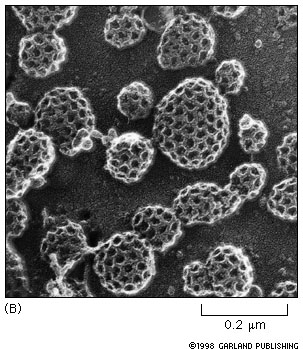
Vesicles form by budding from membranes of ER, Golgi and the
plasma membrane. Micrograph.
Each bud has a distinctive coat protein on cytosol surface.
- The coat protein shapes the membrane into a bud.
- The bud captures the correct molecules for outward transport
because cargo receptors are attached through the membrane to the coat protein
complex. See animation.
- After budding, the protein coat is lost.
- Bud can now interact with target - target interaction signals
are now exposed.
|
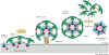
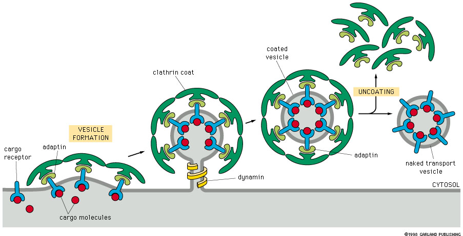
Figure 14-19.
|
Consider endocytosis at the plasma membrane.
Figure 14-19 shows the process of vesicle formation. The same process occurs
at the trans-Golgi to form vesicles that move toward the plasma membrane. |
Fig 14-18 |
Formation of a clathrin coated vesicle. Notice
the thickness of the cargo material attached to the cargo receptors that
extend through the membrane. The clathrins form a layer in the cytosol side
of the membrane. |
This process requires the interaction of several components:
cargo receptor, adaptin, clathrin and dynamin.
- The cargo molecule is picked up by the cargo receptor, which
is an integral membrane protein.
- The cargo receptor/cargo complex is recognized by adaptin
which combines with the cytosolic side of the cargo receptor molecule.
- The cargo/ cargo receptor/ adaptin complex then combines
with clathrin on the cytosolic surface.
- Clathrin forms the curved bud membrane configuration
- Dynamin constricts the neck of the bud (vesicle), which then
pinches off.
- Uncoating then occurs as the clathrin and adaptin are released
and recycled.
- Each vesicle also has a specific targeting signal as described
below.
The vesicle is now ready for transport.
Vesicle targeting
- Over short distances, movement of vesicles is by diffusion.
- Transport of vesicles over longer distances is dependent
on cytoskeleton-based motor proteins.
Docking must be specific. For example, hemicellulose going to
the plant cell wall is delivered to sites where cellulose synthesis is occurring.
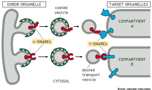
Part of this story involves snares.
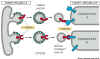
Figure 14-20. |
Snares are proteins that result in specific
attachment of vesicles to their target membranes.
Snares occur as complementary pairs of proteins. The
- vesicle-snare (v-snare) is incorporated into the vesicle
membrane, and the
- target-snare (t-snare) is incorporated into the
target membrane.
- Docking occurs by interaction of the v-snare and t-snare
proteins.
This binding is very specific. This is the part of the targeting process
that corresponds to address on the envelope and house number on the
mail box in the postal delivery analogy.
|
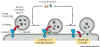
Figure 14-21. |
Once the vesicle and the target membranes are
docked, several other proteins join to form a 'fusion complex' that results
in the fusion of the vesicle with the target membrane. The key protein
here is snap-25 that interacts with the target snare prior to joining of
target and vesicle snares. |
B. Protein Processing and the endomembrane system
All proteins are processed
After translation on ribosomes in the cytosolic compartment
all proteins are processed either in the cytosol or in the ER/Golgi system.
The initial stages of protein processing involve folding.
- Remember that folding of proteins takes place through interaction
with chaperone proteins (see pp 139-40 and 232, 468-9).
- Proteins that are not properly folded are destroyed.
In the cytosol compartment they are tagged with ubiquitin and destroyed by
proteasomes.
Modification of membrane proteins and proteins destined for
secretion in the endoplasmic reticulum. Proteins targeted
to the ER will end up as membrane proteins or as soluble proteins destined for
vesicles (e.g. lysosomal proteins) or secretion.
Other forms of processing occur in the ER lumen.
- covalent modification (phosphorylation,
methylation, acetylation and formation of disulfide bridges), and
- cleavage of the initial protein product to produce
a smaller active protein. Cleavage occurs commonly in the case of digestive
enzymes and other secreted proteins (e.g. insulin).
- glycosylation - addition of polysaccharides to form
glycoproteins.
- The enzymes that carry out these reactions are located in
the lumen of the E R, and not in the cytosol.
- This means that the proteins being glycosylated are bound
for secretion or are membrane proteins.
Thought question: In the case of membrane proteins, what part
of the protein would be glycosylated. The inside (cytosolic) part or the outside
part?
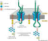 |
Figure 14-22. Protein glycosylation in the
ER. When polypeptide chains enter the endoplasmic reticulum they are immediately
glycosylated by the addition of an oligosaccharide chain that is transferred
as a single unit from a phospholipid called dolichol to an asparagine residue
in the protein |
Note in the figure above (14-22) that the oligosaccharides are
added as an intact pre-fabricated unit consisting of 14 linked sugar residues
transferred from a phospholipid anchor in the membrane.
The glycolipid units are
- initially synthesized in the cytosol and embedded in the
cytosolic face of the membrane. They are then
- flipped to the external (lumen) leaflet of the membrane.
- joined covalently to asparagine in asn -X- (ser or thr) sequence
tag.
C. Control of protein exit from the ER.
Some proteins are retained in the ER (for example, the enzymes
that modify the oligosaccharides that are added to proteins)
- These proteins carry an ER retention signal (KDEL sequence)
at their carboxyl ends. See Table 14-3.
- Even if they get out of the ER into vesicles they are brought
back to the ER by retrograde (trans to cis) movement of transport
vesicles. This is another example of protein targeting via an internal encoded
target signal.
Proteins must be folded and processed properly.
- Proteins that are normally exported from the ER must be properly
folded. Abnormally proteins are retained by chaperone molecules and
degraded if they do not cooperate and fold correctly.
- Many multi-polypeptide proteins, such as antibodies, are
assembled in the ER. If these proteins are not properly assembled (via folding
and the formation of disulfide bridges), the proteins are degraded.
- Cells make lots of mistakes in the assembly of proteins.
They just do not let them be seen in public.
Proteins that get out of the ER are transferred to the Golgi
apparatus by COPII-coated vesicles.
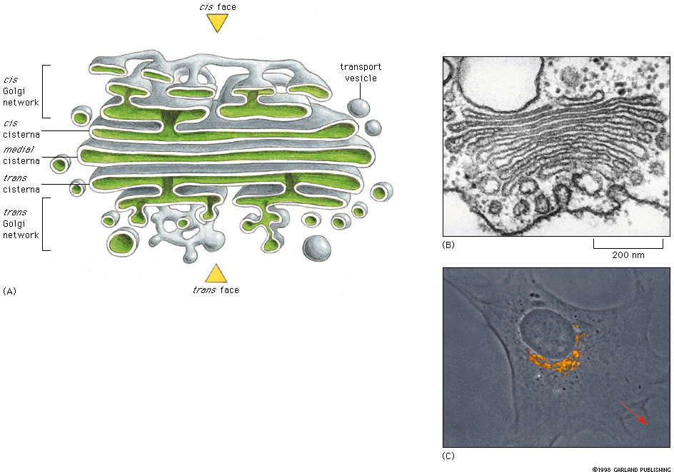
D. The Golgi Complex
Fig 14-24 |
Study Figures 14-24 and 14-17 in text for basic
structure of the organelle. The Golgi complex consists of stack of flattened
sacs (cisternae) with expanded or swollen ends. The Golgi complex has two
functionally and structurally different faces. The behaviour of the Golgi
depends on the presence of other organelles, e.g. cytoskeleton for support
and movement. |
Overview of Golgi
structure.
The Golgi consists of three components:
- the cis Golgi network
- the Golgi stack and
- the trans Golgi network
Each Golgi stack has two faces,
- The cis face, near the ER, is the entry face that
receives small membrane vesicles from the ER. The vesicle membranes are incorporated
into the Golgi membranes and the contents of the vesicles enter the Golgi
cisternae.
- The trans face, facing away from the nucleus toward
the plasma membrane, is the exit face where vesicles leave the Golgi and move
to their targets, including the exterior of the cell.
Here are some images
of Golgi apparatus from the Biol 200 tutorial. Identify
- the cis and trans faces of golgi,
- the trans Golgi network, and
- transport vesicles in these pictures.
The Golgi cisternae contain a variety of transglycosylases (
enzymes that move sugars from one molecule to another) that modify the oligosaccharide
chains of glycoproteins. Different enzymes reside in different regions of the
complex.
The gruesome details of Glycosylation
in the Golgi Complex.
- How are these enzymes kept in place and
- how is the flow of target proteins through the Golgi regulated?
Golgi dynamics:
The flow of cargo proteins through Golgi apparatus is from cis
to trans. (ER > transitional vesicles > cis Golgi Network
> cis cisterna > medial cisterna > trans cisterna > trans Golgi
network > secretory vesicles).
Despite this flow there are many resident proteins that are
localized in particular parts of the Golgi. Two classes of models have been
presented to explain the cis to trans flow of cargo proteins while the resident
proteins stay in place.
1. Vesicle transport model:
- Cargo proteins (but not resident proteins) are moved from
stack to stack by vesicle transport.
- This sorting also involves both
- forward, anterograde (cis to trans),
and
- backward, or retrograde (trans to cis),
flow of vesicles with proteins, moving back up the stack.
- Resident proteins that are carried to locations trans to
their normal location are transported back by retrograde movement of vesicles
2. Cisternal maturation model:
- The cis-most cisterna is the youngest, having been
recently formed from incoming vesicles
- The trans-most cisterna is the oldest and breaks up
into vesicles as material is moved to the trans Golgi network
- Cargo proteins are carried with the cisterna; resident proteins
are returned to their proper location by retrograde movement of vesicles.
There is evidence for both processes, and the extent to
which the actual situation conforms to one model or the other varies among cell
types.
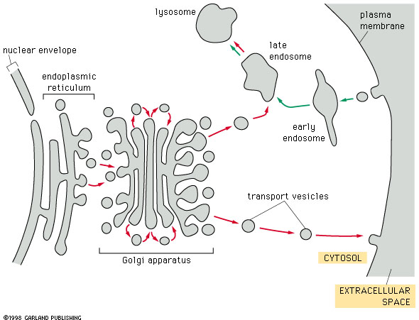
Vesicles from the trans face of the Golgi stack enter
the trans Golgi network, that acts as a sorting and distribution centre.
Vesicles leave the Golgi for a number of destinations. These
include:
- inclusion in lysosomes (for example, enzymes involved in
intracellular digestion),
- incorporation into dense core secretory vesicles that are
stored and later released through the regulated secretory pathway (example,
digestive enzymes in the pancreas) and
- vesicles containing membrane and proteins that are immediately
released to the surface via the constitutive secretory pathway
(example, cell coat proteins).
