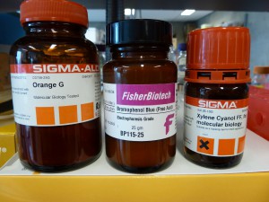
If you are new to the lab bench, and perhaps even if you’re not, you may not know how to make loading dye for agarose gels. Its the sort of thing that tends to get made rarely and then persist, so people often inherit a supply and have no idea how to make more when they run out. For this sort of thing the best reference is the relevant appendix at the back of the Sambrook et al. Molecular Cloning text books (near Megan’s desk) and, of course, there is Google. Or, you can just do what I do. My recipe is incredibly simple and would be close to the cheapest option too.
10X Loading Dye
In an appropriate tube or jar (I usually use a 15ml falcon tube) make a 1:1 mixture of glycerol and whatever buffer you use to make and run your agarose gels, SB for example. Then, add a tiny amount of whatever dye takes your fancy. Start with very little, you can always add more.
So, for example, you could mix 7ml of glycerol and 7ml of SB buffer in a 15ml falcon tube and then add a tiny amount of Orange G dye to make 14ml of an orange 10X loading dye.
A 50/50 glycerol/buffer mixture works well as a 10X loading dye, i.e. it will sink a sample into a well when diluted 10 times, but you can use it at a higher rate if you like. You could treat it as a 6X loading dye if that is what you are used to, (or you could adjust the glycerol/buffer ratio).
I routinely load 15ul volumes when I’m running DNA test gels. I add 2ul of my DNA sample to 11ul of water and 2ul of orange G dyed 10X loading dye. If I had a 20ul PCR that I wanted to run out I’d add 2.5ul of 10X loading dye to it.
Dyes
There are three dyes that are commonly used for this purpose: bromophenol blue, xylene cyanol and orange G. The choice of which dye to use comes down to personal preference based on the speed with which they migrate through agarose and whether you use them as markers to monitor your gels. I don’t use dyes to monitor gels but it is a traditional purpose for a loading dye.
If you use a dye to monitor gels you, obviously, want to use the one you like. Whether you use the dye to monitor your gels or not you need to be careful about quenching the fluorescence of stained DNA (e.g. DNA with EtBr) with an overly dyed gel. This happens when a loading dye has a lot of dye in it, (much more than you actually need to see the sample when loading and to form a visible band of migrating colour in the gel), and that dye migrates at the same speed as a DNA sample of interest. The dye quenches the fluorescent signal from the DNA potentially making DNA of interest difficult to visualise. For this reason its best to not overly dye loading dyes in general but also its a good idea to avoid using a dye that runs at the same speed as DNA of interest.
Bromophenol blue migrates at around 500bp in a 1% gel which is close to the size range of a lot of microsat markers for example. So, if you were running microsats out on agarose the bromophenol blue band in a gel would show you roughly where your markers are, so it would be a good way to monitor the gel, but if it was overly dyed that same band of bromophenol blue would quench the fluorescence you are trying to see.
In a 1% gel:
- xylene cyanol (blue) – around 4000bp.
- bromophenol blue (purple) – around 500bp.
- orange G (orange) – <100bp.
My favourite is orange G. I don’t use the dye to monitor gels so I just want something I can see when I’m loading a gel but that won’t get in the way of anything I want to see fluorescing.
Note that you can combine dyes so that you get more than one band of colour migrating through your gels. A xylene cyanol – bromophenol blue combination is popular.
Dan E.

