STRUCTURE OF THE VERTEBRATE SKULL

Objectives:
1)For the three parts of the skull, be familiar with the following:
-what germ layer it originated from
-location on the skull
-major component bones
-type of bone
-function
2) Be familiar with Williston’s Law
3) Learn the different skull types based on temporal fossae
4) Understand the evolution of the secondary palate
5) Learn how the splanchnocranium contributes to jaw suspension and evolution of the middle ear in vertebrates
COMPOSITION OF THE SKULL
As discussed in lectures there are three elements that contribute to skull formation in the vertebrates:
A) The Neurocranium (Chondrocranium)
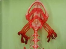
The neurocranium is the portion of the skull that protects the brain and certain sense organs. In the Elasmobranchs (sharks and rays) it is composed of cartilage (chondrocranium), but in most other vertebrates, the cartilage is replaced by bone (endochondral or replacement bone). The neurocranium is a specialized portion of the splanchnocranium and comes from neural crest cells and mesodermal mesenchyme.
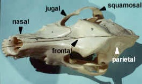
If we think of the entire skull as rectangular box, the bottom, back and sides of the box make up the neurocranium. The bones forming these regions are grouped as the occipitals, sphenoids and ethmoids. The cranial nerves punch through the sides of the box.
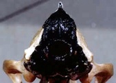
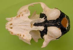
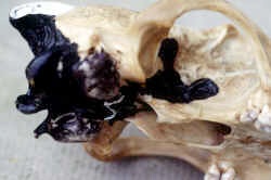
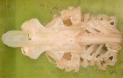
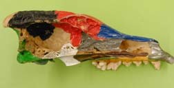
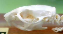
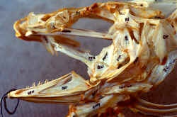
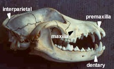
Identify the bones of the neurocranium in the wolf skull provided, using the lab drawings, and table, and the painted skulls on demonstration. Note that these bones are endochondral (or replacement or nondermal) bones. They cannot be distinguished from dermal bone, since they differ only in embryonic origin. The neurocranium bones are painted black on the demonstration wolf and cat skulls.
What group of bones surrounds the opening for the spinal cord (foramen magnum)?
What group of bones has holes for the cranial nerves?
What group of bones forms the back of the box?
What group of bones forms the bottom and sides of the box?
The front of the box is the perpendicular bone dividing the nostrils. What is it called?
On either side, in the nostrils, is a complex of thin bones looking like swirls of flaky pastry. What are they called?
What happens with the original top of the box?
Draw a stylized (rectangular) brain box and label the parts of the neurocranium.
B) The Splanchnocranium
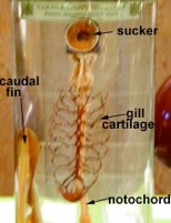
The splanchnocranium consists of the gill arches and their derivatives. The gill arches serve to support the gills and offer a site for respiratory muscle attachment. The original branchial skeleton of cartilage came from neural crest cells.
Splanchnocranium of a cartilaginous dogfish and salamander
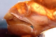
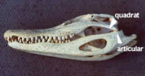
The ossifications of the splanchnocranium of the teleost fishes cannot be readily identified on the wolf skull since they are either the framework for subsequent dermal tooth-bearing bones, or have moved from their association with the jaws to form the ear bones: quadrate (incus), articular (malleus) and hyomandibular (columella or stapes). Look at the alligator and identify the quadrate and articular bones. In mammals, gill arches also form the elements of the larynx (hyoid bone, thyroid and cricoid cartilages). See page 116.
C) The Dermatocranium
The original dermal scales (or armour) of Ostracoderms sink down, attach to the neurocranium and are ossified to form dermal bones. These are from dermatome (epimere mesoderm). The dermatocranium forms most of the skull and functions as a protective shield for the brain.
See the dermal armour of Amia, the bowfin, which sits on the cartilaginous neurocranium. In other animal groups the cartilage disappears and when you look down on the skull you are looking at the bones of the dermatocranium.
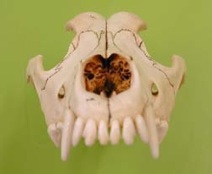
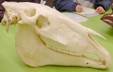
The dermatocranium contributions to the mammal skull that we wish to learn are all those bones that are labeled on the drawing of the wolf skull, plus the dentary bone of the lower jaw. Identify all of these dermal bones on the wolf skull and learn them. These bones are grouped as the facial (upper jaw, nose), vault (front of skull), orbital (around eyes), temporal (side of skull), palatal (roof of mouth) and mandibular (lower jaw) series.
Loss of Dermal Bone
There are skulls of an
alligator and wolf on display. Using the drawings and the skulls provided, identify the dermal bones in each skull. Count the total number of dermal bones (paired (X2) and unpaired (X1) in each species. Please use only the drawings in this lab guide for the count. Note that one pair of bones on one of the drawings is from the splanchnocranium and should not be counted. If any bone is not listed as neurocranium or splanchnocranium on page 39, it is dermal in origin.
The following drawings should provide an understanding of the trend towards loss of dermal bone (Williston’s Law).
Number of dermal bones in Amia skull:
Number of dermal bones in the alligator skull:
Number of dermal bones in wolf skull:
Use the space below to draw the paired wolf dentary bones.
Bones of the skull and their origins
The bones we will concentrate on in this lab are listed here.
Neurocranium (Chondrocranium) is from neural crest cells and mesodermal mesenchyme. It can remain catrilage or become replacement bone. We will study three groups of bones the Occipitals, the Sphenoids and the Ethmoids.
Splanchnocranium comes from neural crest cells and is either cartilage or replacement bone. Arch 1 forms the jaws and is called the mandibular arch. We will particularily study the articular which becomes the malleus of the ear in mammals and the quadrate which forms the incus. Arch 2 is the hyoid arch and we will see the hyomandibular become the collumella and then the stapes of the ear. The hyoid bone also remains as part of the hyoid bone of the larynx. Arches 3-5 are gill arches in fishes and also involved in jaw suspension. They form the caudal portion of the hyoid bone, and the thyroid and cricoid catrilage of the larynx in mammals as seen in the respriation lab on page 116.
Dermal bone is from mesenchyme and ectomesenchyme of the dermis and it overlies the neurocranium and splancnocranium. Learn all the bones on the dorsal drawing of the wolf (pg. 38) plus the lower jaw of mammals (dentary bone) and the new bones, the palatine and bulla.
MODIFICATIONS TO THE SKULL
Temporal Fossae:
Fossae (cavities, pits, or holes), are modifications of the skull that allow for more powerful jaws. They provide more space in the skull for the jaw muscles to expand during contraction and they offer a more secure area for the muscles to attach.
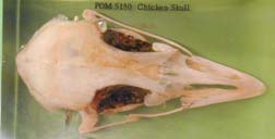
Fish skulls have no fossa and are therefore called anapsid.
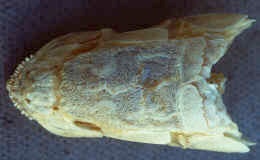
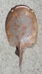
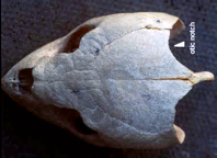
Study the turtle skull on demonstration. See the otic (or temporal) notch in the dorsal posterior region on both sides of the midline. This is an adaptation for muscle attachment that is necessary because of increased jaw musculature, and to offset the interference of the dermal bone contributions. The turtle skull, like the fish skull, has no fossa and is anapsid.
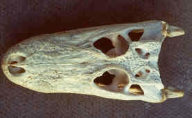
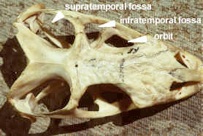
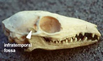
In reptiles (excluding turtles), there evolved a pair of openings on either side of the skull in the temporal region, called the temporal fossa. Study the location of the supratemporal fossa, and the infratemporal fossa on the skull of the alligator. The presence of two temporal fossae is the diapsid condition and is found in some reptiles and birds.
Some fossil reptiles lost the lower (infratemporal) fossa; this is the parapsid (or euryapsid) condition, which is now extinct.
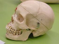
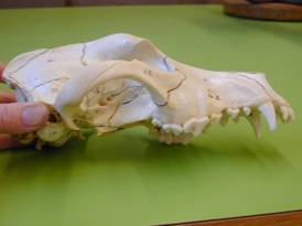
The loss of the supratemporal fossa and the presence of only the infratemporal is the synapsid condition. It occurred in some extinct reptiles, and is represented now by the mammals. Note the eye orbit may be separate from the fossa (cat, horse, human) or confluent with it (wolf, rat).
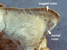
Crests:
Notice on the wolf skull the median longitudinal sagittal crest to attach temporal muscles and a transverse nuchal crest for muscles supporting the head.
THE PALATE
Modifications for breathing air: evolution of the secondary palate. The secondary palate separates the oral passageway from the nasal passageway. There have been three stages in the evolution of the secondary palate:
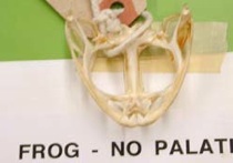
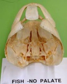
The fishes and amphibia have a complete roof to the mouth which is the primary palate. This is the floor of the neurocranium. This was inconvenient when breathing while eating.
How do adult amphibians, like frogs, get away with eating and breathing at the same time?
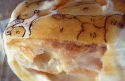
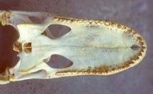
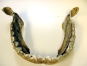
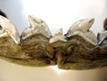
Reptiles show a trend in the evolution of a secondary palate. The turtle on demonstration shows a development of the maxilla, premaxilla, which turn inward to form a shelf, and a new bone, the palatine, which provided a partial secondary palate.
The alligator is a further stage and shows a complete bony secondary palate.
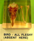
In order to save weight, birds have a totally fleshy secondary palate.
Mammals (wolf, ox) have a functional complete secondary palate, though not the complete bony palate of alligator, the posterior portion being the fleshy soft palate, with the hard palate in the anterior.
What is the functional significance of a secondary palate?
JAW ATTACHMENT TO THE SKULL
Jaw Suspension:
This section explains how the splanchnocranium contributes to jaw attachment to the skull. The mandibular arch (1st segment) of the splanchnocranium formed the upper and lower jaws of cartilage called the palatoquadrate (upper) and Meckel’s cartilage (lower). The hyoid arch (2nd segment) surrounded the spiracle opening.
The Ostracoderms, with one fused head plate of dermal bone (extinct) and modern Agnathans (e.g. Lamprey), lack jaws and display the
condition, in which none of the gill arches are directly associated with the skull. The hyoid arch is a functioning gill arch.
The Placoderms, with several plates of dermal bones in the head (extinct) and primitive fish, Holocephali (e.g. the Rat Fish-Chimaera) display the condition in which the palatoquadrate articulates, or is fused to, the chondrocranium with no supporting function from the hyoid arch. This is the autostylic condition of jaw suspension.
Early sharks and bony fish (now almost all extinct, except for the extant six-gilled shark) had the palatoquadrate attached by ligaments to the chondrocranium. In addition, the hyoid arch specialized to form the hyomandibular, which helped to stabilize the posterior end of the jaws and was attached by a second ligament. This double type of suspension is referred to as amphistylic jaw suspension.
In modern bony fish and modern day sharks (e.g. dogfish) the hyomandibular of the hyoid arch forms a bridge attaching the jaws to the skull. The jaws, free from the skull, can be swung forward a little. Connective tissue and devices like optic pegs help position the jaws. This type of suspension is known as hyostylic. In bony fish, the quadrate and articular bones replaced the cartilage and several dermal bones covered the jaw cartilages. The hyomandibular becomes more firmly attached to the skull and a new bone, the symplectic bone, is formed to aid in jaw movement.
What is the advantage in having a swinging jaw?
In tetrapods, the upper jaw alone suspends the lower jaw. This condition is metautostylic. This frees the hyomandibular of the hyoid arch from jaw suspension and it is incorporated into the ear. The number of upper and lower jawbones becomes reduced. Mammals have only one paired bone, the dentary, in the lower jaw. The articular and quadrate bones are jaw joints in most vertebrates but are moved to the ear in mammals. The entire upper jaw is incorporated into the baincase and jaw suspension is craniostylic.
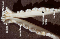
What are the names of the bones of the upper and lower jaws in mammals?
What does the hyomandibular of the hyoid arch do in fishes? Hint: see diagram page 43. Discuss all of the fishes.
What bone does the hyomandibular become in amphibians and reptiles?
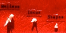
What bone does the hyomandibular become in mammals?
What bone does the articular bone become in mammals?
What bone does the quadrate bone become in mammals?
The three bones of the middle ear and semi-circular canals of the inner ear are enclosed in mammals inside a capsule of new bone, the bulla. Look for this structure in mammals.