LAB 10: THE BRAIN AND EAR

Using your drawings, identify the following brain parts, structures and nerves.
Brain Part Structures Nerves
Telencephalon
olfactory lobes & bulbs,
cerebral hemispheres, ventricles (1&11), part of anterior choroid plexus
olfactory I
Diencephalon
Pineal gland (epiphysis), anterior choroid plexus, optic chiasmia,
Ventricle lll,
neurohypophysis of pituitary gland, thalamus
optic II
Mesencephalon
optic lobes, cerebrial aquiduct
oculomotor III
trochlear IV
Metencephalon
cerebellum, auricles,
Part of 4th ventricle
Myelencephalon
medulla oblongata,
posterior choroid plexus,
posterior part of 4th ventricle
trigeminal V
abducens VI
facial VII
auditory VIII
glossopharyngeal IX
vagus X
NECTURUS BRAIN
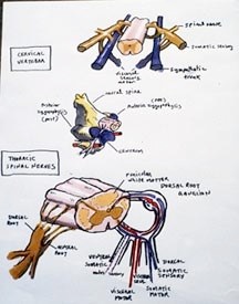
The mudpuppy brain and nerves are similar to that of the dogfish (see previous table). The spinal nerves are more specialized, forming a branchial and lumbrosacral plexus to reflect the addition of limbs.
Remove the skin and muscles from the center of the head. Using forceps and shaving away with a scalpel, pick away the skull until the top of the brain is exposed. Use a dissecting microscope to check how deep you are going. Gently remove the pigmented, vascularized layer and expose as many of the nerves as can be seen dorsally. The nerves follow the pink blood vessels so don’t cut the vessels away. Follow them through the muscles and use your dissecting scope to view the yellow, flat nerves. The ganglia have bone on top of them, which will have to be removed with scalpel and fine forceps.
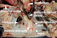
As it was in the dogfish dissection, the inner ear will be exposed. Salamanders do not have an external or middle ear. Read the previous section on the dogfish ear and note the semicircular ducts, and a large otolith in the sacculus.
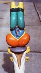
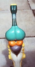
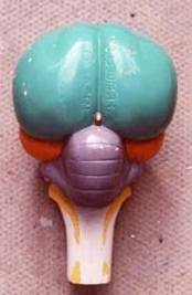
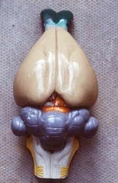
The Telencephalon has olfactory bulbs each with an olfactory nerve, and elongate cerebral hemispheres. The Diencephalon only shows as a small anterior choroid plexus between the posterior part of the cerebral hemispheres and a dorsally projecting pineal gland. The optic nerve II leads to the eye and runs on the outer edge of the sidewall of the brain, along with the ophthalmic nerve, which is just below it. There is usually a pink artery just above both nerves. The Mesencephalon has two rounded optic lobes and oculomotor nerve III and trochlear IV, to the eye muscles. These nerves will not be seen. In the Metencephalon, the cerebellum is poorly developed and is only a narrow transverse band of tissue behind the optic lobes. The Mylencephalon has a large medulla oblongata continuous with the spinal cord. Its parts and nerves are similar to those of the shark. The root of nerves V and VII has a ganglion with bone on top of it. These nerves run in front of the sacculus of the ear, through th muscles, to the upper jaw (maxillary) and the lower jaw (mandiibular), with blood vessels beside them. A short branch of the auditory nerve VIII leads to the sacculus. The root of nerves lX (glossopharyngeal) and X (vagus) has a similar ganglion with bone on top. These nerves run towards the gills behind the sacculus of the ear, with pink blood vessels beside them.
MAMMALIAN BRAIN
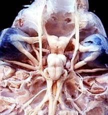
We will be examining whole and bisected sheep brains. Do not cut the whole sheep brains. There are models, keys and photographs on display. The nerves are cut off, so are best seen in the dogfish.
Identify the five parts of the brain and their associated structures.
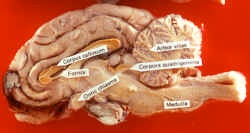
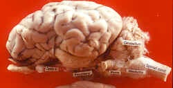
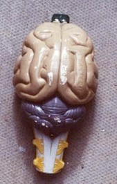
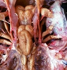
In the Telencephalon, the two large symmetrical structures overlying, and just anterior to, the cerebellum, are the cerebral hemispheres. They are convoluted with hills (gyri) and furrows (sulci) and the grey matter is now on the outside. Within the hemispheres are two cavities; the first and second ventricles joined by the lateral ventricle, which is roofed by the corpus callosum, a commissure to relay information. The olfactory lobes (bulbs) represent the anterior area of the telencephalon. They are two small lobes ventral to the cerebral hemispheres.
The Diencephalon lies between the cerebral hemispheres and the midbrain and is covered by the former. The roof of the diencephalon anteriorly, forms part of the anterior choroid plexus. The cavity of the diencephalon is the third ventricle. The posterior dorsal projection of this part of the brain is the epiphysis or pineal body. This pineal complex is light sensitive; it affects pigmentation, circadian rhythms, reproductive rhythms etc., and synchronizes them with the
external environment. The lateral walls are thalami (thalamus sing.), which are relay and integrating centers for nerve tracts connecting the cerebral hemispheres with the posterior parts of the brain. The hypothalamus, or floor, is ventral, visible externally, and coloured blue in the models. Note the optic chiasma anterior to the hypothalamus. The projection of the hypothalamus is the infundibulum, which is the neurohypophysis (posterior pituitary) from the brain (neural ectoderm). The larger adenohypophysis (Rathke’s pouch), or anterior pituitary, comes from the somatic ectoderm lining of the mouth. This portion often falls off in our preserved sheep brains. Together they form the hypophysis or pituitary gland. The thalamus and pituitary glands affect smooth muscle contraction, membrane permeability, controls reproduction, growth etc., through other glands.
The Mesencephalon is not very visible dorsally, being concealed by the cerebellum, and cerebral hemispheres. The midbrain is painted orange on the models on demonstration. The cavity of the Midbrain is the cerebral aqueduct, or aqueduct of Sylvius, it connects the third ventricle with the fourth ventricle. Locate the optic lobes and a new addition, the auditory lobes. This area is sometimes called the corpora quadragemina as there are now 4 lobes.
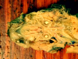
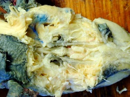
The dorsal portion of the Metencephalon overlies the medulla and is the cerebellum. Its surface is formed into folds and the grey matter has now moved to the outside. It is also called arbor vitae (tree of life). The pons relays information to the cerebral hemispheres.
The most posterior portion of the brain is the large, triangular medulla oblongata of the Mylencephalon. The posterior choroids plexus lies on its dorsal surface. Its posterior limit is the point at which the first pair of spinal nerves emerges. Its cavity is the fourth ventricle. The same spinal nerves emerge as in the shark.
Table of cranial nerves and their function
Cranial nerve
Function in dogfish
Function in mammal
I Olfactory
Sensory. From olfactory bulb to olfactory epithelium. Smell
same
II Optic
Sensory. From optic chiasma to retina. Sight.
same
III Oculomotor
Motor. From floor of mesencephalon to superior rectus, medial rectus, inferior rectus, inferior oblique; and to smooth muscles of iris and ciliary body. Moves eye muscles.
same
IV Trochlear
Motor. From roof of mesencephalon to superior oblique. Moves eye muscle.
same
V Trigeminal
Mixed. From medulla, arising in common with VII and VIII. Sensory to skin of head. Motor to muscles of first gill arch.
Mixed. Sensory to skin of head. Motor to jaw muscles.
VI Abducens
Motor. From medulla to lateral rectus. Moves eye muscle.
same
VII Facial
Mixed. From medulla, arising in common with V and VIII. Sensory to lateral line of head, ampullae of Lorenzini, and mouth. Motor to muscles of second gill arch.
Mixed. Sensory to skin around ear and taste buds of tongue. Motor to muscles of facial expression, lacrymal glands, and mucous glands of oral and nasal cavities.
VIII Auditory
Sensory. From medulla, arising in common with V and VII, to sensory areas of membranous labyrinth.
Sensory to cochlea and vestibular apparatus.
IX Glossopharyngeal
Mixed. From medulla. Sensory to lateral line, first gill pouch, and pharynx. Motor to muscles of third gill arch.
Mixed. Sensory to skin of ear, tympanic membrane, posterior tongue. Motor to tongue and pharynx.
X Vagus
Mixed. From medulla. Sensory to mouth, gill pouches 2-5, and lateral line. Motor to muscles of gill arches 4-7 and cucullaris. Mixed to heart and anterior part of alimentary canal.
Mixed. Sensory to abdominal viscera, esophagus, heart, lungs, bronchi, trachea. Motor to muscles of pharynx and soft palate, trachea, esophagus, larynx, heart, lungs.
XI Accessory
N/A
Motor. From caudal medulla to sternocleidomastoid and trapezius.
XII Hypoglossal
N/A
Motor. From dorsal medulla to muscles to tongue.
SPINAL CORD
The spinal cord is enclosed by the vertebral column and contained in the neural canal; it is continuous with the brain at the foramen magnum. It has grey matter on the inside and white matter on the outside, surrounded by 3 meninges, just like the primitive brain. Spinal nerves of the peripheral nervous system can be seen metamerically arranged along the length of the cord. They exit as dorsal and ventral roots which merge briefly and cross over to emerge as a visceral root to the organs, glands and skin and in a somatic root to the trunk muscles. The visceral (autonomic) nervous system is shown in the diagram. The somatic nerves operate the skeletal and trunk muscles consciously or in a reflex - arc and are responsible for locomotion and movement of the body. Examine the anterior part of the dogfish spinal cord and the models in the lab.
COMPARATIVE BRAIN MODELS
Table: Comparison of brain functions in fish and mammals
LOBE
DOGFISH
MAMMAL
Telencephalon
olfaction and instinctive behaviour
intelligence, memory, instinct (cerebral hemispheres); olfaction.
Diencephalons
integration of sensory and motor
Involuntary actions (emotions, sleep, breathing, temperature), endocrine secretions
Mesencephalon
vision
conscious optic in cerebrum, only reflex optic (i.e. focus) and reflex auditory remain. Motor path to cerebellum via pons. Nerves III & IV.
Metencephalon
integration of balance, hearing and vision (cerebellum)
Integrating locomotion and motor function. Info to cerebellum goes to cerebrum for interpretation, (decision) then back to cerebellum for correlation, relayed by the pons.
Myelencephalon
otic (balance) exit of nerves V-X to gills, jaws, heart.
Otic (hearing and balance) exit of nerves V-X to lungs, heart, digestive tract.
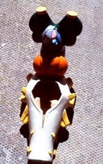
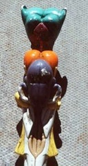
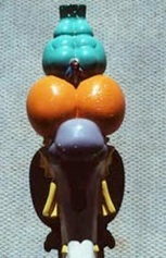
Having reviewed the structure of the dogfish brain, the mudpuppy brain and the dissected sheep brain, examine the series of brain models of dogfish, perch, frog, alligator, pigeon, rabbit and cat, to review the evolutionary story. Note, in particular, that there is little change in the brain until you get to the mammals.
Lamprey Dogfish Perch Frog
Alligator Pigeon Rabbit Cat
Objectives:
1)Learn the structure and function of the ear in each animal.
2)Be able to identify structures and regions of the brain in each animal.
3)Learn the cranial nerves and their function in the dogfish and mammal.
4)Learn the early 3 divisions and later 5 brain divisions in the dogfish.
5)Understand the functions of various regions in the brain.
6)Learn the structural changes in the brain from fish through mammals.
7)Learn the structure of the spinal cord and nerves.
Our study of the nervous system in this lab will be concerned with an examination of the brain of the sheep, and comparison with the dogfish and mudpuppy to show the main evolutionary changes. In the embryo, a three-part brain develops, but adult vertebrates are born with a five-part brain. Examine these five parts in the three animals dissected and note that their structures remain the same. We will also be looking at the ear in the dogfish and a generalized spinal cord.
The brain in encased in membranes called meninges. In dogfish there is only a single meninx, which holds cerebrospinal fluid between it and the brain. The cerebrospinal fluid also circulates within the cavities of the brain and spinal cord. In mudpuppies, there is an outer dura mater and an inner secondary meninx. In mammals, there are three meninges, an outer dura mater, a middle arachnoid membrane and an inner, pia mater.
Table: Divisions of the Brain
3 Part Brain 5 Part Brain
(adult)
(embryonic)
Prosencephalon Telencephalon
(Forebrain) Diencephalon
Mesencephalon Mesencephalon
(Midbrain)
Rhombencephalon Metencephalon
(Hindbrain) Myelencephalon
DOGFISH BRAIN AND EAR
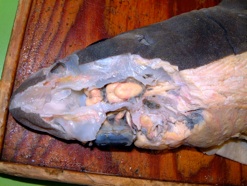
Remove the skin from the dorsal side of the head of your specimen. The cut should be 0.5cm to the left of the mid line to 1.5cm behind the spiracles. Remove the muscles of the head, and in thin horizontal slices, cut away the roof of the chondrocranium. In the area of the eye and inner ear dissect the RIGHT SIDE ONLY. Be careful in this dissection not to cut the cranial nerves, some of which issue dorso-laterally from the brain. As you slice through the hyaline (glassy) cartilage, you will slice through the inner ear. When you have removed the cartilage from the roof and the right side of the head, study the brain in situ.
The Ear
The ear of fishes is an organ of equilibrium, and is specialized for collecting information concerning the position and movements of the head. Although many fish are able to hear, only a very few of the bony fish have a sense of hearing comparable with that of man. The mechanism by which sound vibration is detected is very different in the two groups. In the dogfish the sense of hearing is very poorly developed, and there is no specialization in the structure of the ear for reception of sound, it is primarily done in the lateral line.
To expose the ear requires careful dissection to avoid damaging its delicate structure. To save time, we will slice through the ear in layers while exposing the brain.
Look at the injected plastic models first to get an idea of its size and shape. The diagram below is of the ear from the left side.
The ear of the dogfish lies embedded in the hyaline cartilage of the skull in the otic or auditory capsule.
Locate the spiracle and remove the skin between it and the opening of the endolymphatic duct. Then, with a sharp scalpel, slice fine horizontal slices from the cartilage watching carefully for the first signs of the colourless, worm-like, semi-circular canals. Note the endolymphatic duct, which connects the sacculus of the ear to the outside. Continue cautiously, removing the cartilage around the canals until the whole ear is exposed.
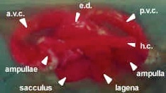
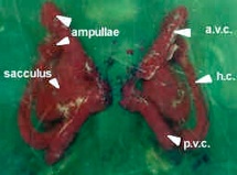
The first structures to appear are the two vertical semicircular canals, the anterior and the posterior. They join at an angle of slightly more than ninety degrees. The horizontal canal projects laterally and horizontally from the ventral ends of the other two.
Examine the figure and note the two ends of the posterior vertical canal are united by the posterior utriculus. The dorsal ends of the anterior vertical and horizontal canals unite to form the anterior utriculus. The latter runs anteriorly and ventrally, and divides to form the ventral ends of the same two canals. Each canal bears a swelling near its ventral end. These are the ampulla, which contains sensitive hair cells in crista (‘c’ in diagram), connecting with a branch of the auditory nerve (cranial nerve VIII).
The main part of the inner ear consists of a large sac-like structure, triangular in lateral view. This is the sacculus. It is connected to each of the utriculi through a pore, and opens to the surface of the head dorsally through the endolymphatic duct. It is usually dark in colour as a result of sand grains, which enter through the endolymphatic duct. Several sensory spots, or maculae (‘m’ in diagram), are located in the walls of the sacculus and utriculi. These contain sensory hair cells (too small to see), which are connected with branches of the auditory nerve (cranial nerve VIII). The nerve branches will be seen later. At the posterior end of the ventral margin of the sacculus is a small out pocketing or depression, the lagena. This is best detected using a blunt probe. In higher vertebrates, this pocket is elaborated to form the cochlea, the part of the ear responsible for the acute sense of hearing.
The semicircular canals, utriculi, and sacculus, which together are known as the membranous labyrinth, contain a fluid, the endolymph that circulates within them. Surrounding the membranous labyrinth and occupying the space between its walls and the cartilage of the chondrocranium is another fluid, the perilymph that prevents the membranous labyrinth from adhering to the chondrocranium.
The sense of equilibrium may be divided into the ability to detect movement of the head, and the ability to detect its position in space. These senses are localized in different parts of the ear. The semicircular canals, ampulla, and utriculi are concerned with the detection of motion. Because of its inertia, the endolymph will tend to circulate around the canals when the head is moved. As the endolymph moves through the ampulla, it bends the delicate hair cells in their walls, and thus initiates nerve impulses to the brain. The plane in which motion of the head occurs will determine which of the six ampullae are stimulated. This is, therefore, an extremely accurate system for providing information concerning the animal’s movement.
The sense of position is centered largely in the sacculus and lagena, which contains sand grains (or, in most vertebrates tiny concretions of calcium carbonate – ‘c’ in diagram). These, known collectively as the otolith, lie free in the endolymph and will alter their position in response to gravity when the position of the head is changed. Depending on their position, they will rest on hair cells in one or other of the maculae, thereby triggering nerve impulses to the brain, and thus providing information concerning the position of the head.
In amphibians, there is a single large, round otolith of calcium carbonate.
In mammals, the sense of equilibrium depends on structures basically similar to those of the inner ear of the dogfish. In addition, modification of the lagena to form the cochlea, and the development of the middle and external ears, has provided man with an extremely sensitive mechanism for detecting sound vibrations. The sense of hearing in the ear is not well developed in the dogfish, and little is known about its mechanism. Possibly sound vibrations in the water impinge on the endolymphatic duct and initiate corresponding vibrations of the endolymph. This in turn might stimulate the maculae.
THE DOGFISH BRAIN
As you slice slivers off the hyaline cartilage, you are slicing through the chondrocranium (neurocranium), a box around the brain. Read the ear section (pgs 136-137) first and look for the parts as you slice. The first part of the brain you will see is the cerebellum, which sticks up; the rest of the brain is deeper. The brain and nerves are cream coloured. Be especially careful of the trochlear nerve, as it runs superficially from the cerebellum through the cartilaginous neurocranium, to the superior oblique muscle of the eye. Try to keep this nerve intact.
Nerves should be teased out of the cartilage with dissecting needles and forceps as the scalpel will cut them off.
The cartilaginous peg (optic peg) between the optic lobe and the eye is the orbital process of the palatoquadrate (upper jaw). It helps keep the jaw in place.