Cable Properties and Action potentials
Chapters
Lodish 4th edition: Chapter 21 pages 923-927
Moyes and Schulte: Chapter 5 pages 168-182
Cable Properties
- properties of the nerve; axon, cell body and dendrite that affect
distance and speed of a membrane potential
- passive conduction properties are called cable because they are analogous to
properties of long copper telecommunication cables (before satellites)
- signal becomes reduced over distance, influenced by cable properties
- loss of current (charge) due to:
Passive flow of current
Signals can be transmitted through a neuron either through passive current flow or using an action potential. Passive flow of current can be thought of in terms of a current travelling down a copper wire.
Figure 21-11. Passive spread of a depolarization of a neuronal plasma membrane with only resting K+ ion channels.
The neuronal membrane is depolarized from -70 to -40 mV at a single point and clamped at this value. The voltage is then measured at various distances from this site. Because of the outward movement of K+ ions through resting K+ channels, the extent of depolarization falls off with distance from the initial depolarization. Passive spread occurs equally in both directions from the site of depolarization. The length constant is the distance over which the magnitude of the depolarization falls to a value of 1/e (e = 2.718) of the initial depolarization. The length constant for a small neuron with a large number of resting K+ channels (black curve) can be as small as 0.1 mm; in this example it is about 0.5 mm. For a large axon, or a small one surrounded by a myelin sheath (blue curve), the length constant can be as large as 5 mm; in this example it is about 1.6 mm.
1) loss of current across membrane (through rest channels)
- loss of current across membrane results in membrane potential dropping with
distance
- dependent on the internal resistance (ri) and the membrane resistance (rm)
- the length or space constant (![]() )
describes this property
)
describes this property
- ![]() = distance (mm) at
which V = 1/e V0 or the distance at which V has decreased to 37%
= distance (mm) at
which V = 1/e V0 or the distance at which V has decreased to 37%
- the relationshipe between the voltage at any distance (x) from the applied
(or original) voltage is :
Vx = Vo e -x/
- ![]() is dependent on
the internal and membrane resistance
is dependent on
the internal and membrane resistance
= square root of (rm/ri)
- if the membrane resistance is large then the longer the impulse will
travel along the nerve before reaching 37% of original
- if the internal resistance is large then the shorter the impulse will travel
along the nerve before reaching 37% of original
- giant axon of squid (1mm dia.)
![]() = 13 mm
= 13 mm
- mammalian nerve fiber (1 micron dia.)
![]() = 0.2 mm
= 0.2 mm
2) loss of current (charge) due to capacitance properties of the
membrane
- cell membrane acts as a capacitor
- 2 conducting sheets separated by an insulating material
- the closer the sheets the better the capacitor
- lipid bilayer is 7 nm thick therefore = excellent capacitor
- it takes time and current (charge) to charge the membrane capacitor
- as current drops over the length of the nerve takes longer and longer to
charge the capacitor
- the time constant describes this effect
- ![]() is the time it takes to
reach 63% of the final voltage (msec)
is the time it takes to
reach 63% of the final voltage (msec)
- ![]() = Rm x Cm
= Rm x Cm
- the smaller the capacitance properties the less the current loss and the
faster the nerve impulse travels
- the large the capacitance properties the more current loss and the slower
the nerve impulse
- time constants range from 1 to 20 msec.
Why are Cable Properties Important to the Function of a Nerve?
Speed of an action potential
- the action potential causes a local depolarization that will passively
spread to adjacent regions
- the neighbouring regions will become depolarized to threshold which will
then trigger an action potential in this region and so on down the length of
the axon
- passive spread of the depolarizing current is the rate limiting step on an
action potential
- if ri is high - current spread is not as far, speed of the action
potential is slower
- if rm is high - internal current spread is farther, speed is
faster
- the velocity of the action potential depends on the rate at which the
membrane capacitance ahead of the action potential can be charged
- this depends on cm and ri
- if cm is high - the longer and more charge it takes to charge the
capacitor and the slower the action potential
Evolutionary Adaptations that Affect Action Potentials
Increased Axon Diameter
- have increased axon diameter in axons to increase action potential
velocity
- i.e. giant axon of squid = 1 mm diameter
why does increasing the diameter of an axon increase the speed of an action
potential?
- rm, ri and cm are all related to the
radius of a fiber
rm ~ 1/2Pradius
ri ~ 1/Pradius2
cm ~ radius
- as increase diameter of a fiber rm and ri decrease, but ri decreases faster,
therefore benefit as the internal resistance decreases faster relative to the
membrane resistance
- therefore the distance the membrane potential can travel is increased by an
increased diameter
- the length constant
![]() is
increased
is
increased
- giant axon of squid (1 mm dia.)
![]() = 13mm
= 13mm
- mammalian nerve fiber (1 micron dia.)
![]() = 0.2mm
= 0.2mm
- increase in fiber diameter also increases cm, but this
increase is proportional to the increase in the radius while the decrease in ri
is proportional to the radius2
- therefore internal resistance decreases faster than the capacitance of the
membrane
- the decrease in ri speeds up the current transfer to the next
region of the nerve and threshold is reached sooner
Myelin
- glial cells called oligodendrocytes and Schwann cells can produce myelin wraps that range from 10-20 up to 160 wraps around the axon
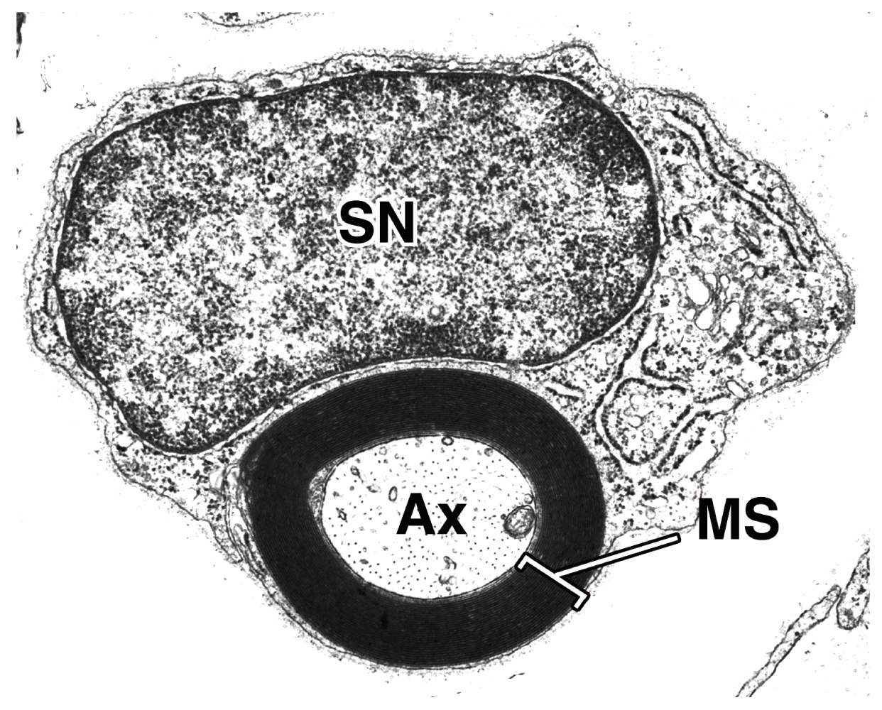
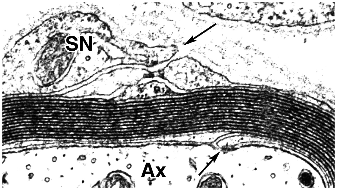
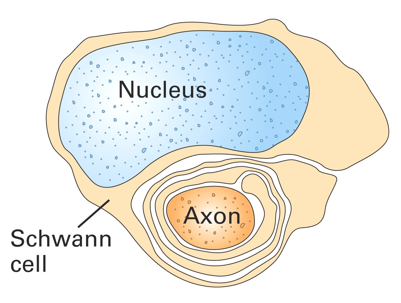
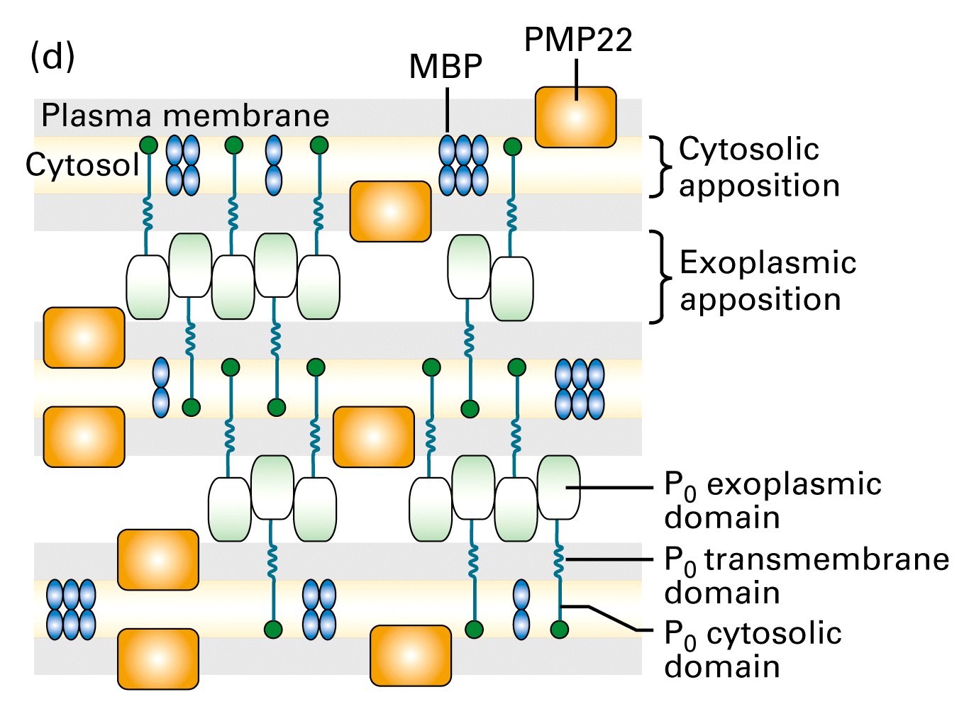
Figure 7-39, Lodish 5th edition. Formation and structure of a myelin sheath in the peripheral nervous system.
Electron micrograph of a cross section of the axon of a myelinated peripheral neuron. It is surrounded by the Schwann cell (SN) that produced the myelin sheath, which can contain 50 100 membrane layers.
As the Schwann cell continues to wrap around the axon, all the spaces between its plasma membranes, both cytosolic and exoplasmic, are reduced. Eventually all cytosol is forced out and a structure of compact stacked plasma membranes is formed.
Composition:
-80 % lipid, 20% protein, (mostly made up of cell membrane)
-during development of myelin the wrappings surround the axon and the
cytoplasm is slowly squeezed out until all that is left is alternating
membranes and a small amount of protein
The compaction of these membranes is generated mainly by a number of proteins
the main being protein zero in the peripheral nervous system which is
synthesized only in myelinating Schwann cells.
Examples to illustrate the effects of myelin wraps on action potential velocity
| Fiber | Myelinated | Fiber diameter | Conduction velocity |
| (mm) | (m/sec) | ||
| A
|
+ myelin | 6 - 12 | 35-75 |
| A
|
+ myelin | 1 - 5 | 5-30 |
| C fibers | - myelin | 0.2-1.5 | 0.5-2 |
- cutaneous C fibers carry pain information, usually a significant delay before you feel pain after burning your finger or hitting it with a hammer
Myelin affects the speed of the action potential
- the action potential causes a local depolarization that will passively spread to the next node of Ranvier to depolarize it to threshold which will then trigger an action potential in this region which will then passively spread to the next node and so on (multiple nodes can be simultaneously depolarized at once)
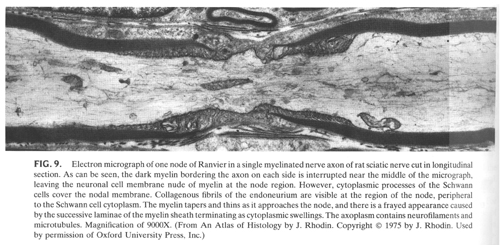
Top: Figure 21-17, Lodish 4th edition. Structure of a peripheral myelinated axon near a node of Ranvier, the gap that separates the portions of the myelin sheath formed by two adjacent Schwann cells. These nodes are the only regions along the axon where the axonal membrane is in direct contact with the extracellular fluid.
Bottom: Figure 23-9, Sperelakis, Cell Physiology, 2nd edition. Electron micrograph of one node of Ranvier in a single myelinated nerve axon of rat sciatic nerve cut in longitudinal section.
- nodes have high concentrations of sodium channels, internodes have very
few (or none)
- therefore more efficient system as fewer Na+ and K+ ions enter and leave the
cell therefore less energy required by the Na-K/ATPase pump to maintain these
concentrations
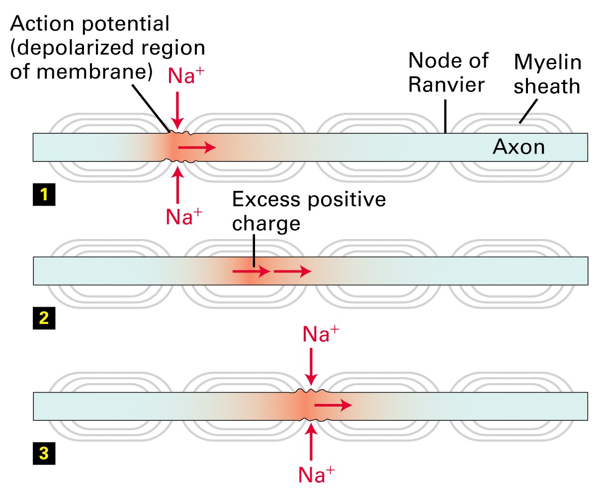
Figure 7-40, Lodish 5th edition. Conduction of action potentials in myelinated axons.
(1) The influx of Na+ ions associated with an action potential at one node results in depolarization of that region of the axonal membrane. (2) Depolarization moves rapidly down the axon because the excess positive ions cannot move outward across the myelinated portion of the axonal membrane. The buildup of these cations causes depolarization at the next node. (3) This depolarization induces an action potential at that node. By this mechanism the action potential jumps from node to node along the axon.
- passive spread of the depolarizing current between the nodes is the rate
limiting step on an action potential
- depends on how much current is lost due the three cable properities
1) if the internal membrane resistance (ri) is high - current spread is not as
far, speed of the action potential is slower
2) if the membrane resistance (rm) is low- current is lost and so current
spread is slower and the action potential slows down
myelin increases rm so that little current is lost,
passive spread of the current is further
3) if the membrane capcitance (cm) is high - the longer and more charge it
takes to charge the capacitor and the slower the action potential
myelin decreases cm so that less current is lost in
charging the capacitor and more is available to spread down the axon