GENERAL FEATURES & FUNCTIONS
Connective tissues are the most abundant of the primary tissues.
They are very different from the epithelial, muscle and nervous tissues.
- In these three tissue types, the cells of the tissue are close together:
 .
.
- In contrast, the cells of the connective tissues are far apart, separated by an
abundant amount of extracellular material, also called extracellular matrix:
 .
.
The properties of the cells and the composition and arrangement of the extracellular
matrix elements vary tremendously, giving rise to an amazing diversity of connective
tissues, each uniquely adapted to perform its specific function in the body.
Where do you find Connective tissues?
everywhere in the body.
What are their functions?
- Binding, support and packaging:
Other tissues (epithelia, muscles, nerves) are supported, surrounded and held in place
by connective tissues. Connective tissue fibers form capsules and membranes which
surround organs, and form ligaments and tendons which bind bones to each other or to muscles.
They also form the 3-dimensional fibrous mesh which supports cells inside large soft organs
such as the liver and spleen. Bone and cartilage support body organs.
The delicate and fragile areolar connective tissue forms a soft packing around organs.
- Protection, defense and repair:
Some connective tissues have great regenerative ability and are important in repair
following injury. Scar tissue is formed of connective tissue which fills in spaces
where the original tissue does not regenerate. Several cellular and molecular
components of connective tissue function in defense against invading bacteria or
chemical substances. Inflammation is a defensive response of connective tissue
at the site of infection or injury. The skull is a bony chamber which protects
the soft brain tissue.
- Insulation:
Fat cells or adipose tissue, is a connective tissue which not only
cushions body organs but also insulates them
and provides reserve energy fuel.
- Transportation:
Blood is a connective tissue and it
carries and delivers oxygen and nutrient to
tissues.
What are the histological characteristics of connective tissues?
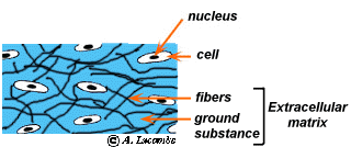
Connective tissues are characterized by abundant amounts of extracellular matrix in which a
variety of cell types are dispersed. The extracellular matrix between the cells usually
includes fibers of one or more types embedded in an amorphous ground
substance.
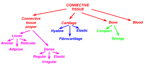
Connective tissues are classified into four classes: BLOOD, BONE,
CARTILAGE, CONNECTIVE TISSUE PROPER.
These four classes of connective tissues are identified on the basis of three criteria:
1- the cell types,
2- the kinds, density and arrangement of their fibers,
3- on the amount and nature of the amorphous ground substance that is present
between its cells.
The cells found in connective tissues can be placed in two categories:
the cells of the connective tissue per see which secrete the matrix or maintain it.
- Each of the major classes of connective tissue contains an undifferentiated cell
type whose name ends in -blast. This cell retains its capacity for division and
secretes the matrix that is characteristic of the tissue. In most connective tissues,
once the matrix is produced, the undifferentiated cells lose their capacity for cell
division and become mature cells whose name ends in -cyste. These mature cells are less
active and in general are responsible for maintaining the matrix in a healthy state.
Fibroblasts are the primary blast cells of the connective tissue proper;
hemocytoblasts are the primary blast cells of the blood; chondroblasts and osteoblasts
are the primary blast cells of cartilage and bone, respectively.
the accessory cells which are supported by the connective tissue.
- Connective tissue (especially one of the proper connective tissues:
the loose connective tissue) is also home to an assortment of other cell types such as:
- fat storing cells that provide reserve energy fuel
- white blood cells; mast cells; macrophages; antibody-producing plasma cells that
are mobile and migrate into the connective tissue matrix from the blood stream.
They are involved in the body defense and in the elimination of dying or dead tissue
cells.
The extracellular matrix is composed of
ground substance and fibers:
- The ground substance
- is the amorphous substance that fills the space between the cells and contains the fibers.
It is composed of interstitial fluid, cell adhesion proteins
and proteoglycans.
Cells adhesion proteins allow the connective tissue cells to attach themselves to
matrix elements. The proteoglycans are proteins to which polysaccharides are attached.
These polysaccharides can trap more or less water depending their nature and form a
substance that varies from a fluid to a semi-stiff hydrated gel. The relative amounts
and kinds of polysaccharides help determine the properties of the matrix.
For example, the more polysaccharides, the stiffer the ground substance is.
The ground substance supports cells, binds them together and functions as a medium through which nutrients and other
dissolved substances can diffuse between capillaries and cells.
- Fibers
- Fibers in the matrix provide strength. Three types of fibers are found in the connective tissue matrix: collagen,
elastic and reticular.
- Collagen fibers (white fibers): are extremely tough. They are stronger
than steel fibers of the same size. They provide high tensile strength, which is the
ability to resist longitudinal stress. Since fresh collagen fibers have a glistening
white appearance they are sometime called "white fibers".
- Elastic fibers (yellow fibers): can be stretched to one and one-half times their length,
but recoil to their initial length when released. They are found where greater elasticity
is needed such as the lungs and the blood vessel walls. Fresh elastic fibers appear yellow
and are also called yellow fibers.
- Reticular fibers: are fine collagenous fibers. They form a delicate branching
network supporting soft organs such as the liver and spleen.
Classification of connective tissues:
There are four classes of connective tissues: BLOOD, BONES, CARTILAGE and
CONNECTIVE TISSUE PROPER. - They are further subdivided into subclasses and types:
I want you to be able to identify all the different types of Connective tissues
as well as learn their locations in the body. Later on we will study bone tissues and blood
in more details.