THE AXIAL AND APPENDICULAR SKELETON EXCLUDING THE SKULL

Objectives:
1) Be able to identify the 3 major types of cartilage and explain where they are prevalent in the body.
2) Be able to differentiate between the 2 major types of bone, describe how they are formed, and where they are located in the body.
3) Identify the additions to vertebrae and their function.
4) Identify the different types of vertebral centra and the vertebrate groups they occur in.
5) Understand regional specialization of vertebrae in the different vertebrate groups.
6) Explain the form and function of the pelvic and pectoral girdles and determine which bones are dermal and which are replacement bones.
7) Identify the major bones in the limbs.
8) Give examples of how limbs are adapted for different functions.
SKELETAL TISSUES
A) Cartilage
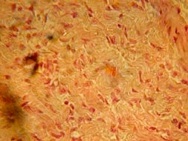
Examine the microscope slides of hyaline cartilage on demonstration. Notice the nuclei of discrete cells in lacunae (spaces) embedded in a matrix of polysaccharides. These cells (chondrocytes) on the underside of the perichondrial membrane secrete the matrix. No blood vessels or nerves penetrate the matrix. All nourishment is by means of diffusion through the matrix, so cartilage seldom acquires much thickness although it grows by internal expansion. Note the different types of cartilage on display. They have different amounts of calcification, which is the addition of calcium salts, and differ in their fibre content. Cartilage is derived from mesenchyme and forms a matrix for bones, ends of bones, intercostals from ribs, nose, ears etc.
Name and draw the three major types of cartilage.
B) General connective tissue
General connective tissue, from embryonic mesenchyme is dispersed throughout the body and also is found as adipose (fat) and areolar tissue and as blood plasma and lymph. Fibrous tissue from the newly discovered syndetome layer (between myotome and sclerotome) makes up tendons, which join bone and muscle together and ligaments, which join bones to each other.
C) Bone
Bone can either be intramembranous (membrane bone) (eg: dermal bone) or replacement.
Dermal bone forms directly in the dermis of the skin from mesenchyme. Osteoblasts form a calcium phosphate matrix and deposit salts, then become osteocytes. Dermal bones are plate like, but can become thicker or grow at the edges. Other membrane bones are deep bones that also do not have a primary cartilage matrix.
Name some dermal bones seen in this week’s lab.
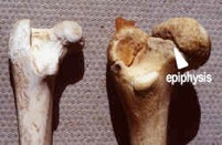
Replacement bones (endochondral bones) are formed by replacing embryonic hyaline cartilage with bone. New cartilage made by chondroblasts grows mainly at the ends and at epiphyses to lengthen the bone. To widen it, the periosteal membrane directly secretes perichondrial bone in strips. Cartilage must be removed before bone can be deposited. The axial skeleton is derived from sclerotome (epimere), the appendicular skeleton from somatic hypomere.
Name some replacement bones seen in this week’s lab.
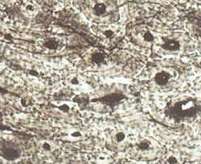
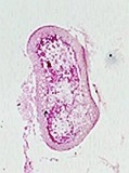
Examine the microscope demonstration of dry ground bone. The forming of this complex structure is called ossification. Refer to the drawing of bone on demonstration and identify Haversian canal systems, (osteons), which are concentric rings of bone surrounding a canal. Identify the canaliculi, lacunae, and bone matrix. The central Haversian canals contain blood vessels, lymphatics and nerves in life, and run longitudinally in the bone. The lacunae contain the bone cells or osteocytes. The lacunae are connected one to another and to the central Haversian canals by small canaliculi and larger Volkmann’s canals. The matrix appears white and is composed of mineral salts, chiefly calcium phosphate.
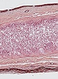
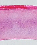
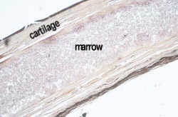
Examine the slides of transverse and longitudinal sections through a long bone (endochondral) in a chick embryo. The sheet of fibrous connective tissue surrounding the outside of the bone, called the periosteum is seen in the chick slides. Compare these slides with the diagrams on demonstration and with the sections through the femur on display.
What is the material in the cavity of living bones and what does it do?
Examine the demonstration material of young and old long bones.
How does a young long bone differ from an older one?
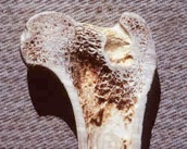
What is the name of the region on either end of a long bone?
What is the name of the shaft?
What is the function of trabeculae?
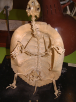
AXIAL SKELETON: VERTEBRAE
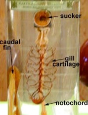
Originally the notochord was the stiffening rod of the phylum Chordata. In the Vertebrata, the notochord is replaced by bony elements of the vertebrae, which are formed by sclerotome. This did not happen all at once, and many early vertebrates show only some vertebral elements, which may be cartilaginous.
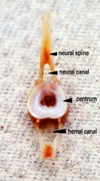
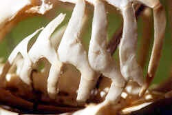
Agnathans, like the lamprey, have tiny cartilaginous neural arches and spines sitting on top of the notochord. Cartilaginous fish like the sharks, have vertebrae of cartilage. At this stage, the elements are all present. The central piece (centrum) is hollow and surrounds the notochord. Dorsally is a neural arch to protect the nerve cord, which is always on top of the notochord. A neural spine extends for muscle attachment. Ventrally, in the tail (caudal) region is a hemal (blood) arch and spine. In the trunk these are sprung open to form ribs. Some early bony fish, like the Sturgeon, lack a centrum and the neural and hemal arches (or ribs) sit directly on the notochord and are of ossified cartilage.
Others, like the bowfin, Amia, show the dispondylous condition, with two vertebrae per myotome. This is only obvious when some of the additions to the centrum are missing. The centrum in Amia contains both a neural and hemal arch (or ribs), whereas the intercentrum lacks these additions in the caudal region. Teleosts are all monospondylous having lost the intercentrum. The vertebrae of Amia and teleosts are completely ossified and the notochord is squeezed to a tiny thread going through a hole in the centrum. Between the centra it enlarges to form a round cushion within the cavities. Amphibia and the rest of the vertebrates have a solid centrum, restriction the notochord to the area between the vertebrae where it contributes to the disks. The shape of these disks varies depending on the positioning of cavities at the anterior or posterior ends of the centrum, either ends or neither end. We shall look at these variations in the ends of the centrum after we look at the additions to the centrum, which together we call the vertebra.
The following translation will be useful in learning the vocabulary of this section.
coel = cavity
amphi = both
pro = in front
opistho = behind
a/an = without
hetero = other
zygos = yoke
pophysis = process
pleur/o = lateral
hypo = below, lower
hyper = upper
dispond = twice
dia = through
para = beside
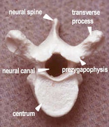
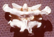
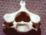
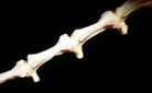
Additions to the Centrum
Caudal Region of a Fish Thoracic Region of a Mammal
Identify the following on the vertebrae provided
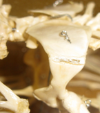
Neural ArchDorsal. What is it protecting?
Neural SpineDorsal. What is attached to it? Look at the skeleton of the cat. Do the spines change direction? Why?
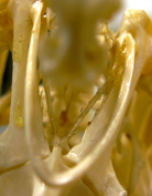
Hemal ArchVentral, found in the caudal region (fish, salamander, alligator). What is it protecting?
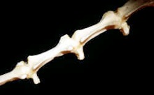
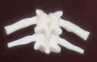
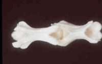
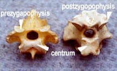
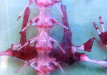
Zygapophyses -Form locking devices between successive vertebrae. They are projections of the dorsal region of the neural arch. Examine Prezygapophyses anteriorly, and Postzygapophyses posteriorly. To distinguish them look only at the articulating surface (the one that rubs) which are called facets. These can be distinguished by the fact that the Prezygapophyses have articulatory surfaces that project anteriorly towards the midline and dorsally, while those of the Postzygapophyses project posteriorly outward and ventrally. Zygapophyses do not occur in the fishes. Zygapophyses prevent torsion and are locked in marine mammals.
Why do marine mammals need more rigid vertebrae?
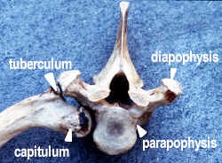
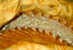
Diapophyses and ParapophysesThey are lateral facets (smooth surfaces), one pair each side of centra in tetrapods, which bear two-headed, ribs. They are not found in fishes or tetrapods with single headed ribs. They articulate with the rib heads. Parapophyses are on the centrum and articulate with the capitulum of the rib. Diapophyses are on the transverse processes, which articulate with the tuberculum of the rib. In what region of the body are they found?
HypopophysesThese are v-shaped midventral projections from the centrum, also known as chevron bones, found only in the anterior caudal region in mammals (e.g. proximal cat tail, opossum tail). What part of the fish vertebrae are they remnants of (i.e. homologous to)?
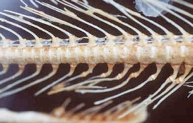
Pleurapophyses These are lateral projections of the centrum, which include short, fused, ribs).
Embryologically, cervical, thoracic, and lumbar ribs are formed. Some reptiles and birds still have cervical and trunk vertebrae. Mammals have cervical pleurapophyses where cervical vertebrae fuse with the embryonic ribs and the hole between the two rib heads becomes the transverse foramen. In the lumbar region, the long transverse processes include fused ribs and so are also termed pleurapophyses.
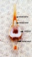
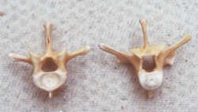
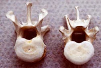
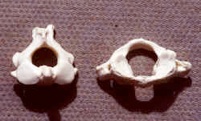
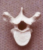
B) Types of vertebrate centra (anterior to left)
Examine the demonstration material on vertebrae types, and identify:
Centra TypeAnimalQuestionAnswer
Amphicoelousfish, salamanderWhere is it concave?
Procoelousalligator, frog, snakeWhere is it concave?
OpisthocoelouslizardWhere is it concave?
AcoelousmammalIs either end concave?
Heterocoelousbird cervical vertebraeHow would you describe the ends?
AXIAL SKELETON: REGIONS
Regional Specialization
Examine the articulated skeletons of fish, amphibian, reptile, bird and mammal that are on demonstration. Notice the following:
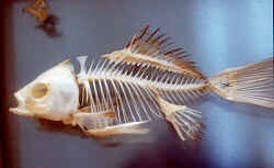
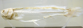
Fish
Two vertebral regions only, trunk, and caudal (tail); trunk vertebrae with ribs, neural arches and spines, caudal vertebrae with neural and hemal arches. In Agnathan (jawless) fish like Ostracoderms and Cyclostomes, the notochord is prominent with small cartilaginous vertebral elements. This condition can also be seen in the sturgeon where the cartilage has been replaced by bone. In sharks and bony fish, the notochord has been reduced to a small thread through the centrum but fills the concavities between vertebrae. Examine the caudal region of the bowfin, Amia and notice that there are two centra per body segment (hypo and pleuro centra). This is the Dispondylous (Diplospondylous) condition. The neural arches, in this caudal region, are borne only on alternate centra. Other fishes display the Dispondylous condition, but duplicate the neural arches only; still others duplicate both arches and centra. Most fishes and higher vertebrates have only one centra per body segment and are Monospondylous.
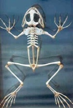
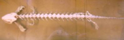
Amphibia
Note four vertebral regions, cervical, trunk, sacral and caudal. The Anurans (tail-less Amphibia) lack the caudal region. Compare the frog and salamander (Necturus) skeletons. Note particularly that both cervical and sacral regions each consist of only one vertebra. The cervical vertebra is the atlas. The atlas is ring-like, and lacks a centrum, and articulates with the occipital condyles of the skull. It enables the head to move up and down but not side to side.
Compare ribs in frog and Necturus. They are fused to the vertebral column in the frogs (pleurapophyses), and are double-headed articulating ribs in Necturus.
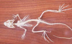
Reptiles
Note five vertebral regions, cervical, thoracic, lumbar, sacral and caudal. Compare the vertebral columns of alligator, snake and turtle. The alligator has 8 cervical, 11 thoracic, 5 lumbar, 2 sacral (fused) and 40 caudal vertebrae. Moveable, double-headed, ribs are borne on the thoracic vertebrae. Ribs, if present on the lumbar vertebrae are fused. Snakes may have as many as 500 vertebrae. Both thoracic and lumbar regions bear ribs. The turtle vertebral column has 8 cervical, 10 trunk, 2 sacral and 16 to 30 caudal vertebrae. The first caudal as well as all the sacral and trunk vertebrae are fused with dermal bone to form the carapace. The ribs are expanded and fused to the inner surface of the costal plates of the carapace. The ribs are single headed. Note that in the Reptiles the two anterior cervical vertebrae are specialized. The first cervical vertebra is called the atlas. The second cervical vertebra, or axis, has an anteriorly projecting process, the odontoid process, which fits into the cavity of the atlas, acting as a pivot in turning the head.
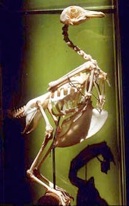
Birds
Rigidity of the vertebral column is achieved by the fusion of many vertebrae. The cervical vertebrae number 13 to as many as 25, and have great flexibility, due to the heterocoelous centra. Note the presence of cervical ribs. There are 5 thoracic vertebrae, but the last one is fused into the synsacrum, and the first four are fused together. The last thoracic, all the lumbar, the 2 sacral and several caudal vertebra all fuse to form one bone, the synsacrum. This in turn is fused to the pelvic girdle. There are several free caudal vertebrae then the tail ends in an enlarged pygostyle, which represents several fused vertebrae. The ribs bear posteriorly projecting uncinate processes, each being ankylosed to the next posterior rib, the distal region of the ribs are joined to the sternum via sternal processes.
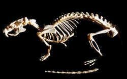
Mammals
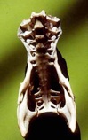
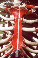
Note the five vertebral regions – cervical (seven vertebrae), thoracic, lumbar, sacral and caudal. In the cat there are 7 cervical, 13 thoracic, 7 lumbar, 3 sacral and 4 to 26 caudal vertebrae. The cervical vertebrae have two holes (foramina) to allow arteries to carry blood to the head. Only the thoracic vertebrae bear mobile ribs. The ribs are double headed. The cervical and lumbar vertebrae bear transverse processes with remnants of ribs fused to them, called pleuropophyses. The three sacral vertebrae fuse to form one bone, the sacrum, with which the pelvic girdle articulates. Between the vertebrae are intervertebral cartilages (disks), which are composed of fibers and notochord remnants.
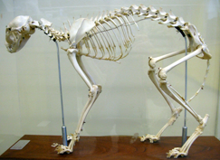
Examine the vertebrae on demonstration and in the cat skeleton. Note the differences between vertebrae from different body regions.
How can you distinguish the atlas?
How can you distinguish the axis?
How can you distinguish a cervical vertebra?
How can you distinguish a thoracic vertebra?
How can you distinguish a lumbar vertebra?
How can you distinguish sacral vertebrae?
How can you distinguish caudal vertebrae?
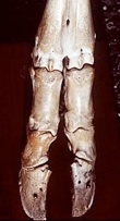
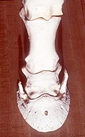
APPENDICULAR SKELETON
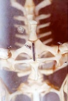
Sternum
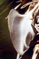
Examine the sternum in the different classes, and notice that in amphibia like salamanders it is of cartilage, while in frogs it is of cartilage and bone and attached to the pectoral girdle only. It acts as a skid and is not attached to the short ribs. Note that in the lizard and alligator the sternum is still cartilaginous and is attached to the ribs. In turtles it is absent but is replaced by the plastron. In the birds, in the flying forms, the sternum is one large bony element, articulating with many ribs. It has a strong central keel, or carina, for insertion of flight muscles.
Examine the sternum on the human skeleton, and notice the rib attachment via the costal cartilages, and the fact that only in mammals is the sternum of many segmented bones.
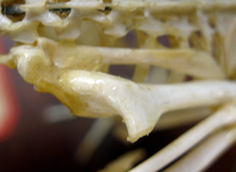
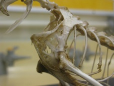
Pectoral Girdle
The pectoral girdle consists of both dermal and endochondral (replacement) bone. The cartilage replacement bones are:
1. Coracoid
2. Scapula
3. Suprascapula
The dermal investing bones are:
1. Clavicle
2. Cleithrum
3. Supracleithrum
4. Post-temporal
Cartilage elements alone occur in the Chondrichthyes (cartilaginous fishes). See the demonstration material of the shark skeleton and pectoral girdles. Note the coracoid bar, lateral scapular processes and the suprascapular processes at their tips. Examine the Teleost (bony fish) pectoral girdle, and note the addition of the dermal elements; the post-temporal bone may abut the posterior of the skull, as in the demonstration. The opercular plates conceal much of the girdle. Only the scapula is ossified in Necturus, and this is correlated with weak limb movement. Examine the demonstration material of the frog pectoral girdle, using the numbered sheet to identify the bones; note the loss of many dermal bones including the cleithrum, associated with movement of the humeral muscles on to the dorsal part of the girdle. The ventral coracoid processes are separate and sometimes overlap.
Frog Sternum and Pectoral Girdle
In the frog, the clavicle is of dermal origin, the sternum is of new origin and the remaining parts are cartilaginous.
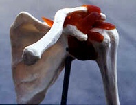
Reptiles have the dermal elements of interclavicles, clavicle and occasionally parts of the cleithrum. Replacement bones are the scapula and a portion of the coracoid (procoracoid). In the Birds, clavicles are retained, and unite to form the furcula (wishbone). Posterior to it runs the paracoracoid bone and a dorsal scapula lies on top of the rib cage. Note loss of the coracoid in Mammals. Mammals have only two bones, the scapula (replacement) and the clavicle (dermal). In running Mammals (cursors) the clavicle may be reduced or absent. See the small splinter of bone in the cat skeleton that is all that remains of the clavicle. Compare this with the prominent clavicle of the Bat and the Opossum. The scapula persists through all the groups, and in the mammals it is a broad flat plate, divided by a scapular spine. It provides the main area for muscle insertion. The small coracoid process is a remnant of the posterior coracoid bone. The scapula articulates with the humerus at the glenoid fossa.
Sketch and label the pectoral girdle of humans.
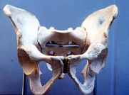
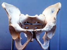
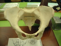
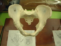
Pelvic Girdle
The pelvic girdle has no dermal elements. It is composed of cartilage replacement bones only. These are the:
1. ILIUM - wing - anterior-dorsal
2. ISCHIUM - posterior -ventral
3. PUBIS - anterior-ventral
Note the simple cartilage girdle in the Shark with a single puboischiac bar and small iliac process near each fin. In the bony fishes it is still simple, but ossified into a single Ischio-pubis, but here, as in the shark, it is still unattached to the vertebral column. Examine the tetrapod pelvic girdles to see the three main ossification areas. In lower forms the ventral area, the puboischiac plate is expanded for limb muscle attachment, but in more advanced forms the ilium becomes the largest element. Note the symphysis that occurs in most forms, but not in the bird. The latter lacks the ventral meeting of elements, and this is associated with the production of a large egg. To maintain strength, the three pelvic bones fuse together as the innominate bone and it is fused with the vertebral synsacrum.
Sketch and label the pelvic girdle in humans.
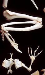
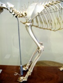
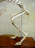
Limbs
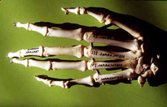
Forelimbs, as early as the amphibians, have a single bone, the humerus in the upper limb and two forearm bones, the larger, anterior radius and posterior ulna, which often articulates with the radius. There are many wrist bones, collectively called the carpals, and the digits are composed of proximal metacarpals and distal phalanges.
Hind limbs consist of an upper limb containing the femur bone and the lower limb with an anterior, larger, tibia and a posterior fibula. The ankle has many bones, collectively called tarsals, and the digits have proximal metatarsals and distal phalanges.
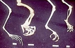
Examine the cat limb for the typical pentadactyl pattern. Compare plantigrade (man) with digitigrade (cat) and with unguligrade (horse). Note the position of the wrist and ankle is quite different in these groups.
How many digits bear weight in plantigrades?
How many digits bear weight in digitigrades?
How do digitigrades differ from plantigrades?
How many digits bear weight in unguligrades?
There are two unguligrade types, mesaxonic, with only one digit (# III) remaining, as found in the horse (perissodactyl, perisso = odd), and paraxonic, with two digits (# III and # IV) remaining to bear the body weight, as found in the pig, cow and deer (artiodactyl).
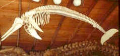
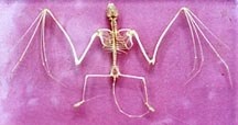
Examine the adaptations for flight and swimming that are on demonstration.